. 2023 Mar 1;11(3):753.
doi: 10.3390/biomedicines11030753.

Cortico-Subcortical White Matter Bundle Changes in Cervical Dystonia and Blepharospasm

Affiliations

Cortico-Subcortical White Matter Bundle Changes in Cervical Dystonia and Blepharospasm

Costanza Giannì et al. Biomedicines. .

Abstract

Dystonia is thought to be a network disorder due to abnormalities in the basal ganglia-thalamo-cortical circuit. We aimed to investigate the white matter (WM) microstructural damage of bundles connecting pre-defined subcortical and cortical regions in cervical dystonia (CD) and blepharospasm (BSP). Thirty-five patients (17 with CD and 18 with BSP) and 17 healthy subjects underwent MRI, including diffusion tensor imaging (DTI). Probabilistic tractography (BedpostX) was performed to reconstruct WM tracts connecting the globus pallidus, putamen and thalamus with the primary motor, primary sensory and supplementary motor cortices. WM tract integrity was evaluated by deriving their DTI metrics. Significant differences in mean, radial and axial diffusivity between CD and HS and between BSP and HS were found in the majority of the reconstructed WM tracts, while no differences were found between the two groups of patients. The observation of abnormalities in DTI metrics of specific WM tracts suggests a diffuse and extensive loss of WM integrity as a common feature of CD and BSP, aligning with the increasing evidence of microstructural damage of several brain regions belonging to specific circuits, such as the basal ganglia-thalamo-cortical circuit, which likely reflects a common pathophysiological mechanism of focal dystonia.

Keywords: basal ganglia-thalamo-cortical circuit; blepharospasm; cervical dystonia; diffusion tensor imaging; focal dystonia; probabilistic tractography; structural MRI; white matter.


Conflict of interest statement

The authors declare no conflict of interest.

Figures

Figure 1
Figure 1
Reconstructed white matter tracts in healthy subjects (HC) ((right) DST patients (left) and BSP patients (center) overlaid on the MNI152 standard brain). Red-yellow colors mean the extent of spatial overlap of reconstructed tracts between participants; specifically, red indicates at least 50% overlap and yellow indicates 100%. Green areas are the regions of interest used for probabilistic tractography.
Figure 2
Figure 2
Differences in fractional anisotropy (FA) of cortical-subcortical white matter tracts among subjects. HS: healthy subjects; CD: cervical dystonia; BSP: blepharospasm; M1: primary motor cortex head/face region; S1: primary sensory cortex head/upper limb region; SMA: supplementary motor area. Kruskal–Wallis post hoc analysis (* p < 0.05). Circles indicates outliers.
Figure 3
Figure 3
Differences in mean diffusivity (MD) of cortical-subcortical white matter tracts among subjects. HS: healthy subjects; CD: cervical dystonia; BSP: blepharospasm; M1: primary motor cortex head/face region; S1: primary sensory cortex head/upper limb region; SMA: supplementary motor area. Kruskal–Wallis post hoc analysis (* p < 0.05, ** p < 0.001). Circles indicates outliers.
Figure 4
Figure 4
Differences in axial diffusivity (AD) of cortical-subcortical white matter tracts among subjects. HS: healthy subjects; CD: cervical dystonia; BSP: blepharospasm; M1: primary motor cortex head/face region; S1: primary sensory cortex head/upper limb region; SMA: supplementary motor area. Kruskal–Wallis post hoc analysis (** p < 0.001). Circles indicates outliers.
Figure 5
Figure 5
Differences in radial diffusivity (RD) of cortical-subcortical white matter tracts among subjects. HS: healthy subjects; CD: cervical dystonia; BSP: blepharospasm; M1: primary motor cortex head/face region; S1: primary sensory cortex head/upper limb region; SMA: supplementary motor area. Kruskal–Wallis post hoc analysis (* p < 0.05, ** p < 0.001). Circles indicates outliers.
Figure 6
Figure 6
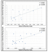
Correlation between diffusion tensor imaging metrics and blepharospasm severity scale is shown. Spearman’s correlation test (r and p are displayed).

References

    1. Conte A., Defazio G., Hallett M., Fabbrini G., Berardelli A. The Role of Sensory Information in the Pathophysiology of Focal Dystonias. Nat. Rev. Neurol. 2019;15:224–233. doi: 10.1038/s41582-019-0137-9. - DOI - PubMed
    1. Conte A., Rocchi L., Latorre A., Belvisi D., Rothwell J.C., Berardelli A. Ten-Year Reflections on the Neurophysiological Abnormalities of Focal Dystonias in Humans. Mov. Disord. 2019;34:1616–1628. doi: 10.1002/mds.27859. - DOI - PubMed
    1. Quartarone A., Hallett M. Emerging Concepts in the Physiological Basis of Dystonia. Mov. Disord. 2013;28:958–967. doi: 10.1002/mds.25532. - DOI - PMC - PubMed
    1. Giannì C., Pasqua G., Ferrazzano G., Tommasin S., De Bartolo M.I., Petsas N., Belvisi D., Conte A., Berardelli A., Pantano P. Focal Dystonia: Functional Connectivity Changes in Cerebellar-Basal Ganglia-Cortical Circuit and Preserved Global Functional Architecture. Neurology. 2022;98:e1499–e1509. doi: 10.1212/WNL.0000000000200022. - DOI - PubMed
    1. Brodoehl S., Wagner F., Prell T., Klingner C., Witte O.W., Günther A. Cause or Effect: Altered Brain and Network Activity in Cervical Dystonia Is Partially Normalized by Botulinum Toxin Treatment. Neuroimage Clin. 2019;22:101792. doi: 10.1016/j.nicl.2019.101792. - DOI - PMC - PubMed

LinkOut - more resources