. 2023 Mar 7;11(3):814.
doi: 10.3390/biomedicines11030814.

Evaluation of Human-Induced Pluripotent Stem Cells Derived from a Patient with Schwartz-Jampel Syndrome Revealed Distinct Hyperexcitability in the Skeletal Muscles

Affiliations

Evaluation of Human-Induced Pluripotent Stem Cells Derived from a Patient with Schwartz-Jampel Syndrome Revealed Distinct Hyperexcitability in the Skeletal Muscles

Yuri Yamashita et al. Biomedicines. .

Abstract

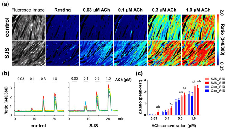
Schwartz-Jampel syndrome (SJS) is an autosomal recessive disorder caused by loss-of-function mutations in heparan sulfate proteoglycan 2 (HSPG2), which encodes the core basement membrane protein perlecan. Myotonia is a major criterion for the diagnosis of SJS; however, its evaluation is based solely on physical examination and can be challenging in neonates and young children. Furthermore, the pathomechanism underlying SJS-related myotonia is not fully understood, and effective treatments for SJS are limited. Here, we established a cellular model of SJS using patient-derived human-induced pluripotent stem cells. This model exhibited hyper-responsiveness to acetylcholine as a result of abnormalities in the perlecan molecule, which were confirmed via comparison of their calcium imaging with calcium imaging of satellite cells derived from Hspg2-/--Tg mice, which exhibit myotonic symptoms similar to SJS symptoms. Therefore, our results confirm the utility of creating cellular models for investigating SJS and their application in evaluating myotonia in clinical cases, while also providing a useful tool for the future screening of SJS therapies.

Keywords: Schwartz–Jampel syndrome; calcium imaging; human-induced pluripotent stem cell; myotonia; perlecan; skeletal muscle.


Conflict of interest statement

The authors declare no conflict of interest.

Figures

Figure 1
Figure 1
Scheme of the differentiation protocol of hiPSCs. PECM: primate ES cell medium, KSR: knockout serum replacement, Lam/En: laminin/entactin, Dox: doxycycline.
Figure 2
Figure 2
Differentiation of control and SJS-derived human-induced pluripotent stem cells (hiPSCs) into myotubular cells. (a) Myosin heavy chain (MHC) staining of myotubes derived from Con_#10, Con_#16, SJS_#10, and SJS_#15; pink shows MHC-positive cells and blue shows nuclei. (b) Differentiation index; the ratio of the number of nuclei in myotubes to the total number of nuclei. (c) The relative RNA expression levels of differentiation markers in skeletal muscle during differentiation. Mean and S.D. are indicated (n  =  3 in each clone). As shown in Figure 1, D0, D2, D4, R2, and R4 correspond to days 0, 2, 4, 6, and 8 after the initiation of differentiation, respectively. UD: undetectable. Data were analyzed using a two-tailed Student’s t-test (b) and one-way ANOVA with Tukey’s multiple comparison (b, c). *: p  <  0.05, **: p <0.01, ***: p  <  0.001, ****: p  <  0.0001. Scale bar, 100 μm.
Figure 3
Figure 3
Perlecan expression in myotubes derived from control and SJS human-induced pluripotent stem cells (hiPSCs). (a) Immunofluorescence analysis of perlecan derived from Con_#10, Con_#16, SJS_#10, and SJS_#15. Green and pink indicate domains III and IV, respectively. (b) The relative RNA expression levels of HSPG2 derived from hiPSCs. (c) The protein expression levels of perlecan extracellularly secreted from hiPSCs. Mean and S.D. are indicated (n  =  3 in each clone). Data were analyzed using a one-way ANOVA with Tukey’s multiple comparison (b,c). *: p  <  0.05, ***: p  <  0.001, ****: p  <  0.0001. Scale bar, 100 μm.
Figure 4
Figure 4
Ca2+ influx in Schwartz–Jampel syndrome (SJS) patient-derived and control human-induced pluripotent stem cell (hiPSC) myotubes. (a) Pseudocolor images of F340/F380 under both resting conditions and peak activity in response to increasing acetylcholine (ACh) concentration (0, 0.03, 0.1, 0.3, and 1.0 µM) in control and SJS-hiPSC myotubes. (b) Representative traces of Ca2+ influx induced by ACh (0, 0.03, 0.1, 0.3, and 1.0 µM) in control and SJS-hiPSC myotubes (n = 5). (c) Dose-response relationship between Ca2+ influx and ACh in control (Con_#10 and #16) and SJS hiPSC myotubes (SJS_#10 and #15). Data were evaluated using the two-way analysis of variance (ANOVA) and Tukey’s multiple comparison test (mean  ±  S.D., n  =  5–6). a. p < 0.05 vs. Con_#10, b. p < 0.05 vs. Con_#16. Scale bar, 100 µm.
Figure 5
Figure 5
Comparison of Ca2+ influx in both perlecan-treated and untreated myotubes derived from Hspg2−/−-Tg mouse satellite cells. (a) Pseudocolor images of changes in F340/F380 at rest and peak stimulation in response to increasing concentrations of acetylcholine (ACh) (0, 0.03, 0.1, 0.3, and 1.0 µM) in both treated (with perlecan; upper panels) and untreated (without perlecan; lower panels) myotubes derived from SJS mice. (b) Representative traces of Ca2+ influx in response to ACh (0, 0.03, 0.1, 0.3, and 1.0 µM) in both treated (with perlecan) and untreated myotubes (n = 5). (c) Dose-response relationship between Ca2+ influx and ACh in both perlecan-treated and perlecan-null myotubes. Data were analyzed using the two-way analysis of variance (ANOVA) and Sidak’s multiple comparison test (mean  ±  S.D., n  =  3). * p < 0.05. Scale bar, 100 µm.

References

    1. Schwartz O., Jampel R.S. Congenital blepharophimosis associated with a unique generalized myopathy. Arch. Ophthalmol. 1962;68:52–57. doi: 10.1001/archopht.1962.00960030056011. - DOI - PubMed
    1. Giedion A., Boltshauser E., Briner J., Eich G., Exner G., Fendel H., Kaufmann L., Steinmann B., Spranger J., Superti-Furga A. Heterogeneity in Schwartz-Jampel chondrodystrophic myotonia. Eur. J. Pediatr. 1997;156:214–223. doi: 10.1007/s004310050587. - DOI - PubMed
    1. Nicole S., Davoine C.S., Topaloglu H., Cattolico L., Barral D., Beighton P., Hamida C.B., Hammouda H., Cruaud C., White P.S., et al. Perlecan, the major proteoglycan of basement membranes, is altered in patients with Schwartz-Jampel syndrome (chondrodystrophic myotonia) Nat. Genet. 2000;26:480–483. doi: 10.1038/82638. - DOI - PubMed
    1. Dagoneau N., Scheffer D., Huber C., Al-Gazali L.I., Di Rocco M., Godard A., Martinovic J., Raas-Rothschild A., Sigaudy S., Unger S., et al. Null leukemia inhibitory factor receptor (LIFR) mutations in Stuve-Wiedemann/Schwartz-Jampel type 2 syndrome. Am. J. Hum. Genet. 2004;74:298–305. doi: 10.1086/381715. - DOI - PMC - PubMed
    1. Arikawa-Hirasawa E., Le A.H., Nishino I., Nonaka I., Ho N.C., Francomano C.A., Govindraj P., Hassell J.R., Devaney J.M., Spranger J., et al. Structural and functional mutations of the perlecan gene cause Schwartz-Jampel syndrome, with myotonic myopathy and chondrodysplasia. Am. J. Hum. Genet. 2002;70:1368–1375. doi: 10.1086/340390. - DOI - PMC - PubMed

LinkOut - more resources