Case Reports
. 2023 Mar 21;11(3):959.
doi: 10.3390/biomedicines11030959.

Co-Occurring X-Linked Agammaglobulinemia and X-Linked Chronic Granulomatous Disease: Two Isolated Pathogenic Variants in One Patient

Affiliations
Case Reports

Co-Occurring X-Linked Agammaglobulinemia and X-Linked Chronic Granulomatous Disease: Two Isolated Pathogenic Variants in One Patient

Lauren Gunderman et al. Biomedicines. .

Abstract

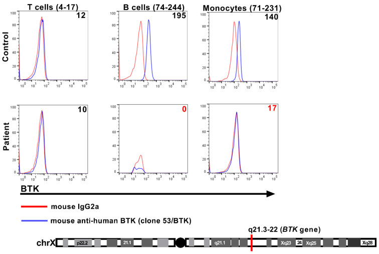
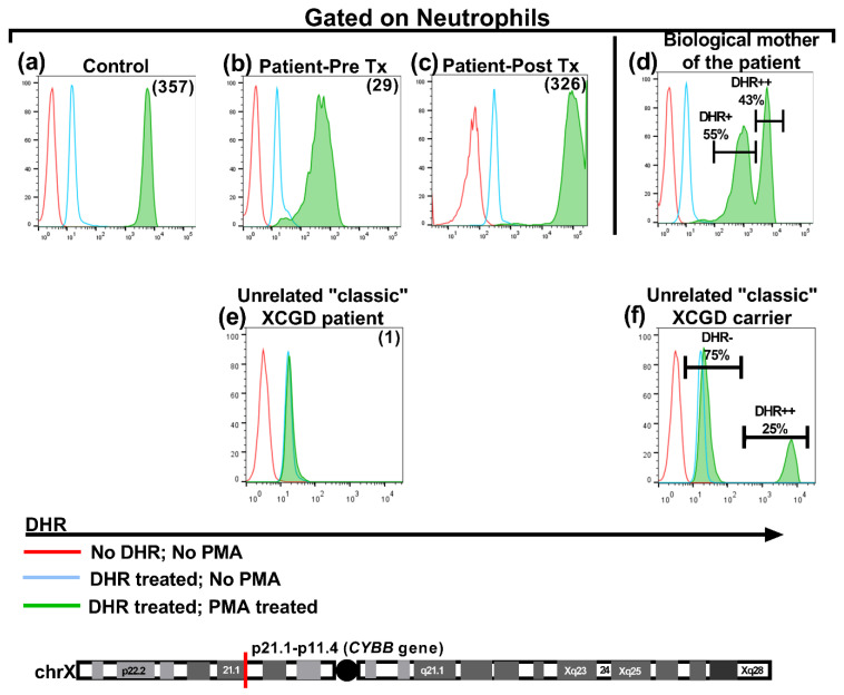
We present a unique and unusual case of a male patient diagnosed with two coexisting and typically unassociated X-linked conditions: he was initially diagnosed with X-linked agammaglobulinemia (XLA) followed by a diagnosis of X-linked chronic granulomatous disease (XCGD) and an as of yet unpublished hypomorphic gp91phox variant in the CYBB gene. The latter was tested after the finding of granulomatous gingivitis. Hematopoietic stem cell transplant (HSCT) was performed due to severe colitis and nodular regenerative hyperplasia (NRH) of the liver. Following transplant, complete donor engraftment was observed with the restoration of a normal oxidative burst and full restoration of normal levels of circulating, mature CD19+ B cells. This case is singular in that it does not involve a contiguous gene syndrome in which deleted genes are in close proximity to either BTK and CYBB, which has been previously reported. To our knowledge, this is the first reported case of XLA and XCGD co-existing in a single patient and of having both inborn errors of immunity successfully treated by HSCT.

Keywords: BTK; HSCT; X-linked agammaglobulinemia; X-linked chronic granulomatous disease; XLA; multi-genetic diagnosis.


Conflict of interest statement

The authors do not have any related conflict of interest.

Figures

Figure 1
Figure 1
Assessment of cytosolic BTK expression by flow cytometry. The histogram overlay plots display the presence and/or absence of BTK protein expression (blue histograms) in the cytosol of the indicated cellular subsets. The numbers listed in parentheses adjacent to the cell subset label reflect the clinically validated reference ranges (5–95th percentiles) for that particular cell subset, derived from the background-subtracted median fluorescence intensity (MFI) of BTK expression evaluated for 34 healthy control donors. The numbers listed within each histogram overlay plot reflect the background-subtracted MFI of BTK expression for that specific cell subset for each donor. The specificity of the BTK signal was determined by also separately staining the cells with a dose-matched, isotype control Ab (red histograms). T cells lack BTK expression and serve as an internal specificity control in the assay.
Figure 2
Figure 2
Histopathological evidence of granulomatous inflammation. (a) Mucosal ulceration with focal granulomatous inflammation, hyperparakeratosis, and acanthosis: image is focused on gingival tissue at the large zone of ulceration. Within fibrous connective tissue, a heavy mixed inflammatory cellular infiltrate that is predominantly neutrophils, lymphocytes, and histiocytes is seen. Widely scattered ill-defined clusters of histiocytes and multinucleated giant cells are noted. (b) Biopsy of transverse colon tissue showing large bowel mucosa with few granulomas. No significant crypt distortion. (c) Biopsy of the liver performed via needle biopsy. Hepatic architecture shows subtle distortion with nodular areas of widened alternating with areas of narrowed liver cell plates. No significant portal inflammation or interface hepatitis. Many of the portal areas lack a distinctive portal vein, and there are occasional foci of lobular inflammation. There is mild focal steatosis, involving less than 5% of parenchyma.
Figure 3
Figure 3
Assessment of neutrophil oxidative burst activity by flow cytometry. Three aliquots of whole blood from each donor were utilized to evaluate neutrophil oxidative burst activity. The first aliquot was left untreated. The second aliquot was labeled with dihydrorhodamine (DHR) and left unstimulated. The third aliquot was labeled with DHR and stimulated with phorbol 12-myristate 13-acetate (PMA) to induce neutrophil oxidative burst activity. The numerical values listed in parentheses in the overlaid histogram plots depict the neutrophil oxidative index (NOI), which is derived by dividing the median fluorescence intensity (MFI) of Rhodamine 123 observed in the PMA-stimulated tube (third aliquot) by the MFI of DHR measured in the second aliquot (unstimulated tube) (normal NOI >30). The DHR fluorescence patterns for the healthy control (a) and the patient sample pre- and post-stem cell transplant (b,c) are displayed. The DHR fluorescence in the biological mother’s sample (d) displays a “variant” bimodal distribution XCGD carrier pattern on account of the hypomorphic CYBB variant and random inactivation of the X-chromosome (lyonization). The frequencies of the maternal neutrophils harboring the normal (wild-type) and variant (mutated) CYBB genes are listed adjacent to the appropriate histogram peaks (DHR++ and DHR+). As a point of reference, the DHR fluorescence patterns from an unrelated case of “classic” XCGD (e) and a “classic” XCGD carrier (f) are also displayed. The frequencies of the neutrophils harboring the normal (wild-type) and mutated CYBB gene for the classic XCGD carrier are listed adjacent to the appropriate histogram peaks (DHR++ and DHR-) Tx: transplant.

References

    1. Chan K.-W., Wong C.-Y., Leung D., Yang X., Fok S.F.S., Mak P.H.S., Yao L., Ma W., Mao H., Zhao X., et al. Targeted Gene Sanger Sequencing Should Remain the First-Tier Genetic Test for Children Suspected to Have the Five Common X-Linked Inborn Errors of Immunity. Front. Immunol. 2022;13:883446. doi: 10.3389/fimmu.2022.883446. - DOI - PMC - PubMed
    1. El-Sayed Z.A., Abramova I., Aldave J.C., Al-Herz W., Bezrodnik L., Boukari R., Bousfiha A.A., Cancrini C., Condino-Neto A., Dbaibo G., et al. X-linked agammaglobulinemia (XLA): Phenotype, diagnosis, and therapeutic challenges around the world. World Allergy Organ. J. 2019;12:100018. doi: 10.1016/j.waojou.2019.100018. - DOI - PMC - PubMed
    1. O’Toole D., Groth D., Wright H., Bonilla F.A., Fuleihan R.L., Cunningham-Rundles C., Sullivan K.E., Ochs H.D., Marsh R., Feuille E. X-Linked Agammaglobulinemia: Infection Frequency and Infection-Related Mortality in the USIDNET Registry. J. Clin. Immunol. 2022;42:827–836. doi: 10.1007/s10875-022-01237-1. - DOI - PMC - PubMed
    1. Marciano B.E., Spalding C., Fitzgerald A., Mann D., Brown T., Osgood S., Yockey L., Darnell D.N., Barnhart L., Daub J., et al. Common severe infections in chronic granulomatous disease. Clinical infectious diseases: An official publication of the Infectious Diseases Society of America. Clinical Infectious Diseases. 2015;60:1176–1183. doi: 10.1093/cid/ciu1154. - DOI - PMC - PubMed
    1. Oikonomopoulou Z., Shulman S., Mets M., Katz B. Chronic Granulomatous Disease: An Updated Experience, with Emphasis on Newly Recognized Features. J. Clin. Immunol. 2022;42:1411–1419. doi: 10.1007/s10875-022-01294-6. - DOI - PMC - PubMed

Publication types

LinkOut - more resources