Review
. 2023 Mar 22;11(3):990.
doi: 10.3390/biomedicines11030990.

Structure-Guided Prediction of the Functional Impact of DCLK1 Mutations on Tumorigenesis

Affiliations
Review

Structure-Guided Prediction of the Functional Impact of DCLK1 Mutations on Tumorigenesis

Annalisa L E Carli et al. Biomedicines. .

Abstract

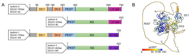
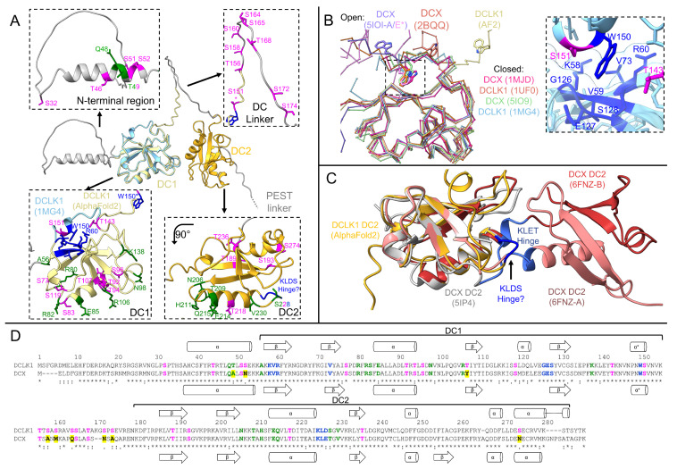
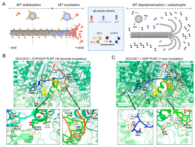
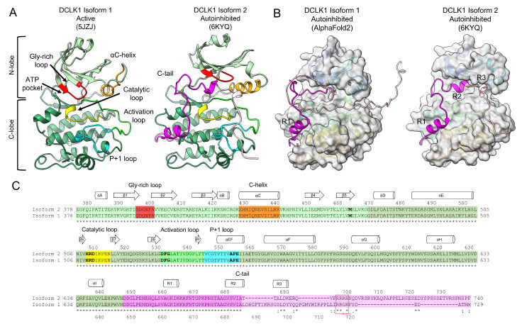
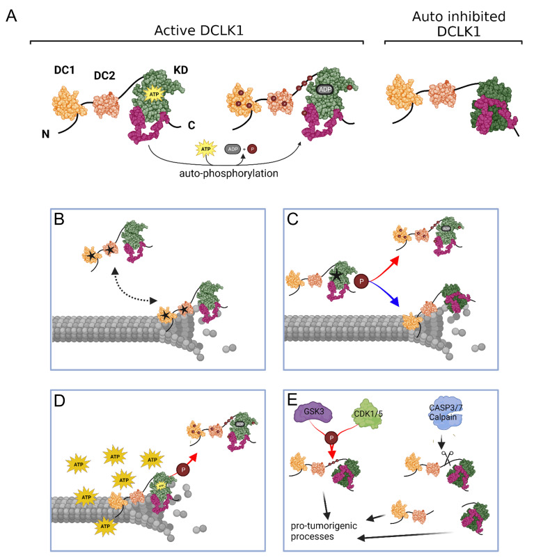
Doublecortin-like kinase 1 (DCLK1) is a functional serine/threonine (S/T)-kinase and a member of the doublecortin family of proteins which are characterized by their ability to bind to microtubules (MTs). DCLK1 is a proposed cancer driver gene, and its upregulation is associated with poor overall survival in several solid cancer types. However, how DCLK1 associates with MTs and how its kinase function contributes to pro-tumorigenic processes is poorly understood. This review builds on structural models to propose not only the specific functions of the domains but also attempts to predict the impact of individual somatic missense mutations on DCLK1 functions. Somatic missense mutations in DCLK1 are most frequently located within the N-terminal MT binding region and likely impact on the ability of DCLK1 to bind to αβ-tubulin and to polymerize and stabilize MTs. Moreover, the MT binding affinity of DCLK1 is negatively regulated by its auto-phosphorylation, and therefore mutations that affect kinase activity are predicted to indirectly alter MT dynamics. The emerging picture portrays DCLK1 as an MT-associated protein whose interactions with tubulin heterodimers and MTs are tightly controlled processes which, when disrupted, may confer pro-tumorigenic properties.

Keywords: DCLK1; DCX; PEST domain; cancer; cryo-EM; crystal structure; doublecortin domain; kinase; microtubules; missense mutations.


Conflict of interest statement

The authors declare no conflict of interest.

Figures

Figure 1
Figure 1
Doublecortin-like kinase 1 (DCLK1) structure and domain organization: (A) schematic overview of the four DCLK1 isoforms and domain boundaries comprising the two tandem doublecortin (DC) domains (yellow/orange), the proline, glutamic acid, serine, and threonine rich (PEST) linker-region (light blue), the S/T-kinase domain (KD, light green), and the short (S, purple) and long (L, magenta) regulatory C-terminal tail (C-tail); and (B) cartoon representation of the AlphaFold2 model of DCLK1 (Isoform 2, O15075) colored according to confidence (Predicted Local Distance Difference Test—pLDDT) (AlphaFold2 [63,64]). Regions with a pLDDT value of < 50 are likely disordered and the positions of corresponding amino acids relative to domains are uncertain. Structure rendered in UCSF ChimeraX v1.5 [65].
Figure 3
Figure 3
Mapping of residue function on the DC domains of DCLK1: (A) cartoon representations of the AlphaFold2 prediction of DCLK1 (Isoform 2, yellow/gray) and the crystal structure of DCLK1-DC1 (PDB ID: 1MG4, light blue). Zoom panels highlight functional residues in each domain: αβ-tubulin binding (green), conformational switch (blue), and auto-phosphorylation (magenta); (B) comparison of open and closed structures of DC1 from DCLK1 and DCX, showing only the C-alpha backbone for clarity. PDB IDs are provided in parentheses. The zoom panel shows the tryptophan (W)150 pocket in DCLK1 (PDB ID: 1MG4) with surrounding residues (blue) highlighting nearby phosphorylation sites (magenta); (C) comparison of monomeric structures of DC2 with the domain swapped dimer structure (salmon/red). The KLET hinge in DCX which flips out to mediate the domain swap, and the equivalent residues in DCLK1 (KLDS), are highlighted in blue; and (D) sequence alignment of DCLK1 and DCX colored according to assigned function as in (A). Differences in functional residues are highlighted in yellow. Experimentally determined and predicted secondary structures are depicted. Residue numbers for DCLK1 are shown above with the boundaries of the DC domains indicated. Sequence alignment performed using PROMALS3D [84] and structures rendered in UCSF ChimeraX v1.5 [65].
Figure 2
Figure 2
Structural basis of microtubule (MT) stabilization and polymerization by doublecortin protein (DCX): (A) schematic depiction of DCX domain functions. The more flexible doublecortin 2 (DC2) domain is involved in MT nucleation and preferably binds guanosine triphosphate (GTP)-αβ tubulin at the growing plus ends of the MTs. The more rigid DC1 domain binds guanosine diphosphate (GDP)-αβ tubulin and stabilizes MTs and protects them from MT catastrophe. Upon phosphorylation, both DC domains dissociate from the MTs resulting in MT depolymerization/catastrophe; and (B,C) time-resolved cryo-electron microscopy (EM) structures of DCX bound to MTs ((B) PDB ID: 6RF2/(C) PDB ID: 6REV [73]). Each DC domain is depicted in a cartoon representation (rainbow color from N- (blue) to C-terminus (red)). DCX residues which form hydrogen bonds or salt bridges with tubulin (green) are displayed as sticks and labeled in bold with the equivalent residue in DCLK1 in parentheses. Zoom panels highlight key interfaces. Distances for hydrogen bonds are indicated (orange). Schematic generated with BioRender.com and structures rendered in UCSF ChimeraX v1.5 [65].
Figure 4
Figure 4
The structure and conformations of the DCLK1 kinase domain: (A) cartoon representation of DCLK1 structures in the active conformation (left, isoform 1, PDB ID: 5JZJ) and autoinhibited conformation (right, isoform 2, PDB ID: 6KYQ), with the N-lobe (light green) and C-lobe (olive green) indicated. Catalytic and regulatory regions are highlighted: the glycine rich loop (red), αC-helix (orange), the catalytic loop (yellow), the activation loop (green), the P + 1 loop (cyan), and the C-tail (purple/pink); (B) comparison of the C-tail structure predicted by AlphaFold2 [63,64] for isoform 1, and the auto-inhibited structure of isoform 2. The kinase domains are depicted in a surface representation excluding the C-tail portion. The three regulatory helices of the C-tail (R1, R2, R3) are indicated; and (C) sequence alignment of DCLK1 isoforms 1 and 2 colored according to assigned function as in (A). Key functional motifs are bolded: the gatekeeper residue (M465), HRD-motif in the catalytic loop, DFG-motif in the activation loop, and the APE-motif in the P + 1 loop. Secondary structure is shown for isoform 2. The Arginine (R) rich region in the C-tail is partially conserved (dashed box). Sequence alignment performed using PROMALS3D [84] and structures rendered in UCSF ChimeraX v1.5 [65].
Figure 5
Figure 5
Schematic summary depicting how DCLK1 mutations can affect MT dynamics: (A) the active and autoinhibited form of DCLK1: (B) mutations within the DC domains can interfere with MT binding, affecting MT polymerization and stability; (C) as can mutations which increase or decrease kinase activity; (D) increased kinase activity can result due to excess adenosine triphosphate (ATP) in cancer cells; and (E) additionally, regulation by upstream kinases or cleaved products can lead to activation of pro-tumorigenic processes. Figure created with BioRender.com.

Similar articles

Cited by

References

    1. Bateman A., Martin M.J., Orchard S., Magrane M., Agivetova R., Ahmad S., Alpi E., Bowler-Barnett E.H., Britto R., Bursteinas B., et al. UniProt: The Universal Protein Knowledgebase in 2021. Nucleic Acids Res. 2021;49:D480–D489. doi: 10.1093/NAR/GKAA1100. - DOI - PMC - PubMed
    1. Moores C.A., Perderiset M., Kappeler C., Kain S., Drummond D., Perkins S.J., Chelly J., Cross R., Houdusse A., Francis F. Distinct Roles of Doublecortin Modulating the Microtubule Cytoskeleton. EMBO J. 2006;25:4448–4457. doi: 10.1038/sj.emboj.7601335. - DOI - PMC - PubMed
    1. Akhmanova A., Steinmetz M.O. Control of Microtubule Organization and Dynamics: Two Ends in the Limelight. Nat. Rev. Mol. Cell Biol. 2015;16:711–726. doi: 10.1038/nrm4084. - DOI - PubMed
    1. Mohan R., John A. Microtubule-Associated Proteins as Direct Crosslinkers of Actin Filaments and Microtubules. IUBMB Life. 2015;67:395–403. doi: 10.1002/iub.1384. - DOI - PubMed
    1. Sossey-Alaoui K., Srivastava A.K. DCAMKL1, a Brain-Specific Transmembrane Protein on 13q12.3 That Is Similar to Doublecortin (DCX) Genomics. 1999;56:121–126. doi: 10.1006/geno.1998.5718. - DOI - PubMed

LinkOut - more resources