. 2023 Mar 16;12(6):912.
doi: 10.3390/cells12060912.

Mitochondrial Haemoglobin Is Upregulated with Hypoxia in Skeletal Muscle and Has a Conserved Interaction with ATP Synthase and Inhibitory Factor 1

Affiliations

Mitochondrial Haemoglobin Is Upregulated with Hypoxia in Skeletal Muscle and Has a Conserved Interaction with ATP Synthase and Inhibitory Factor 1

Brad Ebanks et al. Cells. .

Abstract

The globin protein superfamily has diverse functions. Haemoglobin has been found in non-erythroid locations, including within the mitochondria. Using co-immunoprecipitation and in silico methods, we investigated the interaction of mitochondrial haemoglobin with ATP synthase and its associated proteins, including inhibitory factor 1 (IF1). We measured the expression of mitochondrial haemoglobin in response to hypoxia. In vitro and in silico evidence of interactions between mitochondrial haemoglobin and ATP synthase were found, and we report upregulated mitochondrial haemoglobin expression in response to hypoxia within skeletal muscle tissue. Our observations indicate that mitochondrial pH and ATP synthase activity are implicated in the mitochondrial haemoglobin response to hypoxia.

Keywords: ATP synthase; ATP synthase inhibitory factor 1; haemoglobin; hypoxia; mitochondria.


Conflict of interest statement

The authors declare no conflict of interest.

Figures

Figure 1
Figure 1
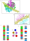
Molecular docking of IF1 and tetrameric haemoglobin, and a visualisation of interacting residues. (a) Docking of tetrameric haemoglobin (red) with a single chain of IF1; (b) the interacting residues of IF1 in haemoglobin docking simulation; (c) schematic representation of interacting residues between haemoglobin chain A and IF1 chain; (d) positioning of IF1 in proximity to interacting haemoglobin chains A and C, where different coloured lines represent different interactions between amino acids, specifically: red—salt bridges, yellow—disulphide bonds, blue—hydrogen bonds, and orange—non-bonded contacts; (e) schematic representation of the interacting residues between haemoglobin chain C and IF1 chain.
Figure 2
Figure 2
Molecular docking of IF1 and monomeric haemoglobin alpha, and a visualisation of interacting residues. (a) Docking of monomeric haemoglobin alpha (green) with a single chain of IF1; (b) the monomeric haemoglobin alpha interaction site of IF1 chain; (c) schematic representation of the interacting residues between monomeric haemoglobin alpha and IF1 chain.
Figure 3
Figure 3
Haemoglobin–IF1 association after 8 ns MD simulation. (a) IF1 docked in the cleft of two alpha chains of haemoglobin (pink—Chain A, light blue—Chain C, yellow—IF1). (b) Shows interaction of haemoglobin residues with IF1 residues—Serine 84 and Asparagine 131 of HbA. Interacting residues. (c) Schematic representation of interacting residues between tetrameric haemoglobin chain A and IF1 after 8 ns MD simulation. (d) Positioning of IF1 in proximity to interacting haemoglobin chains A and C after 8 ns MD simulation, where different coloured lines represent different interactions between amino acids, specifically: red—salt bridges, yellow—disulphide bonds, blue—hydrogen bonds, and orange—non-bonded contacts. (e) Schematic representation of interacting residues between tetrameric haemoglobin chain C and IF1 after 8 ns MD simulation.
Figure 4
Figure 4
Quantitative quality check for the MD run. (A) Root-mean-square deviation (RMSD); (B) radius of gyration shows a reasonable invariant Rg values across the MD run of 8 ns, indicating the protein remains very stable; (C) the thermodynamic factors of density, pressure, and temperature are stable across the MD run.
Figure 5
Figure 5
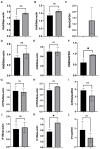
Relative mitochondrial protein expression in normoxic (black) and hypoxic (grey) (A,D,G,J) rat liver,(B,E,H,K) mouse liver, and (C,F,I,L) mouse quadriceps muscle. (AC) haemoglobin α; (DF) haemoglobin β; (GI) ATP5A; (JL) IF1. Liver mitochondria protein expression normalised to β-actin and quadriceps muscle mitochondria protein expression normalised to GAPDH (see Supplementary Figure S4). Data presented as mean with SEM, * p < 0.05, N = 3, Student’s unpaired t-test.
Figure 6
Figure 6
Relative expression of mitochondrial proteins in normoxic and hypoxic D. melanogaster. (A) haemoglobin; (B) IF1; (C) ATP5A. Protein expression normalised to β-actin (see Supplementary Figure S5). Data are presented as mean with SEM, * p < 0.05, ** p < 0.005 N = 3, Student’s unpaired t-test.
Figure 7
Figure 7
Atractyloside-treated HEPG2 cells show a trend toward increased expression of mitochondrial haemoglobin. (A) Relative expression of haemoglobin α in mitochondria isolated from atractyloside-treated HEPG2 cells. (B) Relative expression of haemoglobin β in mitochondria isolated from atractyloside-treated HEPG2 cells. Data are presented as mean with SEM, N = 3, student’s unpaired t-test.

References

    1. Perutz M.F., Rossmann M.G., Cullis A.F., Muirhead H., Will G., North A.C.T. Structure of Hæmoglobin: A three-dimensional fourier synthesis at 5.5-Å. resolution, obtained by X-ray analysis. Nature. 1960;185:416–422. doi: 10.1038/185416a0. - DOI - PubMed
    1. Benesch R., Benesch R.E. The effect of organic phosphates from the human erythrocyte on the allosteric properties of hemoglobin. Biochem. Biophys. Res. Commun. 1967;26:162–167. doi: 10.1016/0006-291X(67)90228-8. - DOI - PubMed
    1. Benesch R., Benesch R.E., Yu C.I. Reciprocal binding of oxygen and diphosphoglycerate by human hemoglobin. Proc. Natl. Acad. Sci. USA. 1968;59:526–532. doi: 10.1073/pnas.59.2.526. - DOI - PMC - PubMed
    1. Greaney G.S., Hobish M.K., Powers D.A. The effects of temperature and pH on the binding of ATP to carp (Cyprinus carpio) deoxyhemoglobin. J. Biol. Chem. 1980;255:445–453. doi: 10.1016/S0021-9258(19)86194-X. - DOI - PubMed
    1. Thomas C., Lumb A.B. Physiology of haemoglobin. Contin. Educ. Anaesth. Crit. Care Pain. 2012;12:251–256. doi: 10.1093/bjaceaccp/mks025. - DOI

Publication types

MeSH terms

Substances

LinkOut - more resources