Review
. 2023 Mar 20;12(6):946.
doi: 10.3390/cells12060946.

Cytological Diagnosis of Classic Myeloproliferative Neoplasms at the Age of Molecular Biology

Affiliations
Review

Cytological Diagnosis of Classic Myeloproliferative Neoplasms at the Age of Molecular Biology

Sophie Combaluzier et al. Cells. .

Abstract

Myeloproliferative neoplasms (MPN) are clonal hematopoietic stem cell-derived disorders characterized by uncontrolled proliferation of differentiated myeloid cells. Two main groups of MPN, BCR::ABL1-positive (Chronic Myeloid Leukemia) and BCR::ABL1-negative (Polycythemia Vera, Essential Thrombocytosis, Primary Myelofibrosis) are distinguished. For many years, cytomorphologic and histologic features were the only proof of MPN and attempted to distinguish the different entities of the subgroup BCR::ABL1-negative MPN. World Health Organization (WHO) classification of myeloid neoplasms evolves over the years and increasingly considers molecular abnormalities to prove the clonal hematopoiesis. In addition to morphological clues, the detection of JAK2, MPL and CALR mutations are considered driver events belonging to the major diagnostic criteria of BCR::ABL1-negative MPN. This highlights the preponderant place of molecular features in the MPN diagnosis. Moreover, the advent of next-generation sequencing (NGS) allowed the identification of additional somatic mutations involved in clonal hematopoiesis and playing a role in the prognosis of MPN. Nowadays, careful cytomorphology and molecular biology are inseparable and complementary to provide a specific diagnosis and to permit the best follow-up of these diseases.

Keywords: cytomorphology; laboratory practice; molecular biology; myeloproliferative neoplasms.


Conflict of interest statement

The authors declare no conflict of interest.

Figures

Figure 1
Figure 1
Bone marrow biopsy of Polycythemia Vera. Panmyelosis with an increase in pleomorphic mature megakaryocytes. (optical microscopy, May Grünwald Giemsa (MGG), low magnification ×15).
Figure 2
Figure 2
Indirect signs of myelofibrosis on blood smear. (a) Leukoerythroblastosis with a polychromatophilic erythroblast cell (green arrow) and immature granulocytic cell (orange arrow); (b) Teardrop-shaped RBC (black arrow) and basophilic cell (blue arrow). (Optical microscopy, May Grünwald Giemsa, ×50 magnification).
Figure 3
Figure 3
Diagnostic algorithm for PV.
Figure 4
Figure 4
Cytological aspects of megakaryocytes in ET BM compared to physiological BM. (a) Physiological megakaryocyte in BM; (b) Cluster formation of megakaryocytes in BM ET; (c,d) Giant megakaryocytes and hypersegmented nuclei with «staghorn-like» aspect in ET BM. (optical microscopy, MGG and 100× magnification).
Figure 5
Figure 5
Blood smear in pre-PMF. Heterogeneity of platelets with macroplatelet (red arrows), giant platelet (purple arrow), degranulated macroplatelet (grey arrow) associated with teardrop-shaped RBC (black arrow). (Optical microscopy, May Grünwald Giemsa, ×100 magnification).
Figure 6
Figure 6
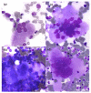
Morphological aspect of megakaryocytes in BM pre-PMF. (a) Atypical megakaryocytes with variable size (large to small) in pre-PMF: with hyperchromatic nuclei; (bd) Enlarged megakaryocytes with variable lobulation; (e,f) Scattered small megakaryocytes with nude nuclei in platelet clusters. (Optical microscopy, May Grünwald Giemsa, ×100 magnification).
Figure 7
Figure 7
Bone marrow cellularity and morphological aspects of megakaryocytes in Chronic Myeloid Leukemia. (a) Granulocytic hyperplasia and small MKs (pink arrows) (optical microscopy, May Grünwald Giemsa (MGG), ×20 magnification) (b) A small MK and eosinophil (yellow arrows) and basophil (blue arrows) cells (optical microscopy, May Grünwald Giemsa (MGG), ×50 magnification).
Figure 8
Figure 8
Algorithm diagnosis for CML, ET, prePMF.

References

    1. Shallis R.M., Wang R., Davidoff A., Ma X., Podoltsev N.A., Zeidan A.M. Epidemiology of the classical myeloproliferative neoplasms: The four corners of an expansive and complex map. Blood Rev. 2020;42:100706. doi: 10.1016/j.blre.2020.100706. - DOI - PubMed
    1. Goldman J.M. Chronic myeloid leukemia: A historical perspective. Semin. Hematol. 2010;47:302–311. doi: 10.1053/j.seminhematol.2010.07.001. - DOI - PubMed
    1. Cortes J., Pavlovsky C., Saußele S. Chronic myeloid leukaemia. Lancet. 2021;398:1914–1926. doi: 10.1016/S0140-6736(21)01204-6. - DOI - PubMed
    1. Sawyers C.L., McLaughlin J., Witte O.N. Genetic Requirement for Ras in the Transformation of Fibroblasts and Hematopoietic Cells by the Bcr-Abl Oncogene. J. Exp. Med. 1995;181:307–313. doi: 10.1084/jem.181.1.307. - DOI - PMC - PubMed
    1. Steelman L.S., Abrams S.L., Whelan J., Bertrand F.E., Ludwig D.E., Bäsecke J., Libra M., Stivala F., Milella M., Tafuri A., et al. Contributions of the Raf/MEK/ERK, PI3K/PTEN/Akt/mTOR and Jak/STAT pathways to leukemia. Leukemia. 2008;22:686–707. doi: 10.1038/leu.2008.26. - DOI - PubMed

MeSH terms