Review
. 2023 Mar 22;12(6):964.
doi: 10.3390/cells12060964.

Adult and Pediatric Nail Unit Melanoma: Epidemiology, Diagnosis, and Treatment

Affiliations
Review

Adult and Pediatric Nail Unit Melanoma: Epidemiology, Diagnosis, and Treatment

Jade Conway et al. Cells. .

Abstract

Nail unit melanoma (NUM) is an uncommon form of melanoma and is often diagnosed at later stages. Approximately two-thirds of NUMs are present clinically as longitudinal melanonychia, but longitudinal melanonychia has a broad differential diagnosis. Clinical examination and dermoscopy are valuable for identifying nail findings concerning malignancy, but a biopsy with histopathology is necessary to confirm a diagnosis of NUM. Surgical treatment options for NUM include en bloc excision, digit amputation, and Mohs micrographic surgery. Newer treatments for advanced NUM include targeted and immune systemic therapies. NUM in pediatric patients is extremely rare and diagnosis is challenging since both qualitative and quantitative parameters have only been studied in adults. There is currently no consensus on management in children; for less concerning melanonychia, some physicians recommend close follow-up. However, some dermatologists argue that the "wait and see" approach can cause delayed diagnosis. This article serves to enhance the familiarity of NUM by highlighting its etiology, clinical presentations, diagnosis, and treatment options in both adults and children.

Keywords: acral melanoma; longitudinal melanonychia; nail; nail unit melanoma; pediatric melanoma; subungual melanoma.


Conflict of interest statement

Conway has no conflicts of interest. Bellet has no conflicts of interest. Rubin has no conflicts of interest. Lipner has served as a consultant for Orth-Dermatologics, Hoth Therapeutics, and BelleTorus Corporation.

Figures
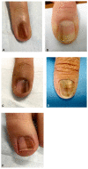
Figure 1
Figure 1
Clinical appearance of subungual melanoma cases. (A), The patient is a 26-year-old female with a 5-mm brown band (68% width percentage) on the nail of the right ring finger. (B), The patient is a 63-year-old woman with a 2.5-mm brown band (18.6%) on the right thumbnail. (C), A 42-year-old man with a 6-mm brown band (54.5%) on the nail of the left fifth finger. (D), A 52-year-old woman with a 7.5-mm brown band (60.0%) on the left thumbnail. (F), An 18-year-old woman with a 5.5 mm dark brown band (46.4%) on the nail of the left hallux. Reproduced with permission from ref. [19].
Figure 2
Figure 2
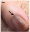
Invasive melanoma in a band of LLE. Dermoscopy highlights splinter hemorrhages and distal triangular onycholysis. Reproduced with permission from ref. [20].
Figure 3
Figure 3
A Rare Case of Osteoinvasive Amelanotic Melanoma of the Nail Unit; clinical photo demonstrating a hypervascular, nonpigmented lesion on the dorsum right distal hallux. Reproduced with permission from ref. [21].
Figure 4
Figure 4
Nail unit melanoma clinical features—LM and Hutchinson sign (arrows). Reproduced with permission from ref. [3].
Figure 5
Figure 5
Dermoscopic appearance of subungual melanomas. (B), Brown lines on a brown background, irregular color, thickness, and spacing with no loss of parallelism. (C), Brown lines on a brown background, irregular color, thickness, and spacing with loss of parallelism. Reproduced with permission from ref. [19].
Figure 6
Figure 6
(A,D,F), Nail unit melanoma dermoscopic features—brown bands irregular in color, width, and spacing, and Hutchinson sign (arrows). Reproduced with permission from ref. [3].
Figure 7
Figure 7
Histopathology of in situ matrix melanomas. Hematoxylin and eosin stain. (A), Almost invisible changes. (B), A few cells are obvious (arrow). (C), The entire matrix epithelium shows pagetoid spread of atypical melanocytes (arrows). (D), Lentiginous (small arrows) and nest-like (large arrow) proliferation of atypical melanocytes. Reproduced with permission from ref. [3].
Figure 8
Figure 8
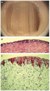
Representative clinical morphology, photomicrographs of H&E staining, and IHC staining for cyclin D1 and PRAME of selected subungual melanoma in situ. One 44-year-old patient (A), 2 mm-wide melanonychia showed (B), atypical melanocyte proliferation with confluency and pagetoid spread (200× magnification, H&E). Reproduced with permission from ref. [42].
Figure 9
Figure 9
Nail unit melanoma after surgical outcome—good aesthetic and functional outcome. (A,B,D,E) defects required full-thickness skin graft for closure. (C,F), were allowed to heal secondarily. Reproduced with permission from ref. [3].
Figure 10
Figure 10
Subungual melanoma in situ (subungual lentiginous melanocytic proliferation with atypia). (A), A 13-year-old girl was referred to complete treatment of a lesion diagnosed as subungual melanoma in situ. The clinical examination revealed the nail bed to show no signs of persistence of the tumor after complete excision of the nail unit. (B), Panoramic view of a transverse biopsy of the matrix showing subepidermal fissures and intraepithelial cell proliferation. (C), Greater detail (×200) shows a lentiginous proliferation of atypical melanocytes, with the formation of fissures between the epithelium and underlying dermis and the focal suprabasal ascent of melanocytes, which completely replaces the keratinocytes in the basal layer. (D), High magnification (×400) reveals the atypical cellular characteristics of the proliferation: large melanocytes with pyknotic and pleomorphic nuclei and suprabasal ascent in some areas. Reproduced with permission from ref. [75].
Figure 11
Figure 11
13-year-old girl presented with a painless black macule affecting her left thumbnail, later diagnosed as subungual melanoma. Clinical features of melanonychia in this case. (A), A homogenous black macule involving 70% of the nail plate, with a hypomelanotic lesion of proximal nail plate and irregular pigmentation extended to the hyponychium. (B), Dermoscopic features (polarized light) of the distal nail plate and hyponychium. Reproduced with permission from ref. [76].
Figure 12
Figure 12
(A), 6-month-old boy with a band of longitudinal melanonychia of the first right toe. Dermatoscopy shows a dark-brown longitudinal band with lines exhibiting irregular coloration, spacing, and thickness. (B), Low-power view of the heavily pigmented matrix lesion. (C), High-power view of the mainly lentiginous proliferation of large, atypical melanocytes. Reproduced with permission from ref. [77].
Figure 13
Figure 13
(A), 11-year-old girl with a longitudinal melanonychia of the second right fingernail. Dermatoscopy shows a pale brown background with lines of irregular coloration, spacing, and thickness. (B), Scanning view of large irregularly spaced melanocytes. (C), MelanA stain reveals abnormal positive melanocytes. Reproduced with permission from ref. [77].

References

    1. Bradford P.T., Goldstein A.M., McMaster M.L., Tucker M.A. Acral lentiginous melanoma: Incidence and survival patterns in the United States, 1986–2005. Arch. Dermatol. 2009;145:427–434. doi: 10.1001/archdermatol.2008.609. - DOI - PMC - PubMed
    1. Basurto-Lozada P., Molina-Aguilar C., Castaneda-Garcia C., Vázquez-Cruz M.E., Garcia-Salinas O.I., Álvarez-Cano A., Martínez-Said H., Roldán-Marín R., Adams D.J., Possik P.A., et al. Acral lentiginous melanoma: Basic facts, biological characteristics and research perspectives of an understudied disease. Pigment. Cell Melanoma Res. 2021;34:59–71. doi: 10.1111/pcmr.12885. - DOI - PMC - PubMed
    1. Duarte A.F., Correia O., Barros A.M., Ventura F., Haneke E. Nail melanoma in situ: Clinical, dermoscopic, pathologic clues, and steps for minimally invasive treatment. Dermatol. Surg. 2015;41:59–68. doi: 10.1097/DSS.0000000000000243. - DOI - PubMed
    1. Kazi R., Moghaddam S., Chu P., Marghoob A.A. Histologic Evidence of Melanocytes Isolated to the Nail Matrix. JAMA Dermatol. 2016;152:573–575. doi: 10.1001/jamadermatol.2015.5869. - DOI - PubMed
    1. Lee D.J.R., Arbache S.T., Quaresma M.V., Nico M.M.S., Gabbi T.V.B. Nail Apparatus Melanoma: Experience of 10 Years in a Single Institution. Ski. Appendage Disord. 2018;5:20–26. doi: 10.1159/000488722. - DOI - PMC - PubMed