

Contrast Enhanced Mammography (CEM) Enhancing Asymmetry: Single-Center First Case Analysis
- PMID: 36980319
- PMCID: PMC10047777
- DOI: 10.3390/diagnostics13061011
Contrast Enhanced Mammography (CEM) Enhancing Asymmetry: Single-Center First Case Analysis
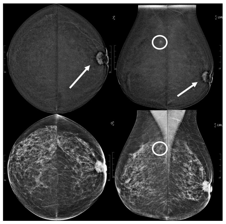
Abstract
(1) Purpose: The latest Breast Imaging Reporting and Data System (BI-RADS) lexicon for CEM introduced a new descriptor, enhancing asymmetries (EAs). The purpose of this study was to determine which types of lesions were correlated with EAs. (2) Methods: A total of 3359 CEM exams, executed at AOUC Careggi in Florence, Italy between 2019 and 2021 were retrospectively assessed by two radiologists. For each of the EAs found, the size, the enhancing conspicuity (degree of enhancement relative to background described as low, moderate, or high), whether there was a corresponding finding in the traditional radiology images (US or mammography), the biopsy results when performed including any follow-up exams, and the presence of background parenchymal enhancement (BPE) of the normal breast tissue (minimal, mild, moderate, marked) were described. (3) Results: A total of 64 women were included, 36 of them underwent CEM for a preoperative staging assessment, and 28 for a problem-solving examination. Among the 64 EAs, 19/64 (29.69%) resulted in being category B5 (B5) lesions, 5/64 (7.81%) as category B3 (B3) lesions, and 40/64(62.50%) were negative or benign either after biopsy or second-look exams or follow-up. We assessed that EAs with higher enhancing conspicuity correlated significantly with a higher risk of B5 lesions (p: 0.0071), especially bigger ones (p: 0.0274). Conclusions: EAs can relate both with benign and tumoral lesions, and they need to be assessed as the other CEM descriptors, with re-evaluation of low-energy images and second-look exams, particularly larger EAs with higher enhancing conspicuity.
Keywords: CEM; breast cancer; breast imaging; enhancing asymmetry; second look.
Conflict of interest statement
The authors declare no conflict of interest.
Figures


References
-
- Bicchierai G., Busoni S., Tortoli P., Bettarini S., Di Naro F., De Benedetto D., Savi E., Bellini C., Miele V., Nori J. Single Center Evaluation of Comparative Breast Radiation dose of Contrast Enhanced Digital Mammography (CEDM), Digital Mammography (DM) and Digital Breast Tomosynthesis (DBT) Acad. Radiol. 2022;29:1342–1349. doi: 10.1016/j.acra.2021.12.022. - DOI - PubMed
LinkOut - more resources
Full Text Sources
