

The Importance of Immunohistochemistry in the Evaluation of Tumor Depth of Primary Cutaneous Melanoma
- PMID: 36980327
- PMCID: PMC10046945
- DOI: 10.3390/diagnostics13061020
The Importance of Immunohistochemistry in the Evaluation of Tumor Depth of Primary Cutaneous Melanoma
Abstract
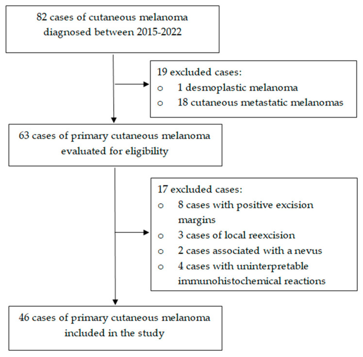
Primary cutaneous melanoma (PCM) is the most aggressive skin malignancy, with an increasing incidence and significant mortality. Tumoral invasion, expressed as Breslow thickness, is routinely assessed on hematoxylin and eosin (HE), although this stain may sometimes underestimate the tumoral depth. The aim of this study was to compare the efficiency of the immunohistochemical (IHC) markers S-100, SOX10, Melan-A, and HMB-45 with HE for the evaluation of the Breslow thickness and staging of PCM. This retrospective study included 46 cases of PCM diagnosed between 2015 and 2022; for each case, the Breslow thickness using HE, S-100, SOX10, Melan-A, and HMB-45 was measured and the appropriate T category was recorded. The highest values of the Breslow thickness were observed for S-100. However, S-100, SOX10, and Melan-A provided statistically significant higher values of the Breslow thickness compared to HE, but no difference was noted between HMB-45 and HE. S-100 was most frequently involved in increasing the T category (26.1%), the majority of cases being upstaged from T1a to T1b. The IHC markers S-100, SOX10, and Melan-A contributed to better evaluation of the melanoma invasion, especially in thin melanomas, but their impact on staging and consecutive treatment remains to be confirmed by future studies.
Keywords: Breslow thickness; immunohistochemical markers; primary cutaneous melanoma; tumor staging.
Conflict of interest statement
The authors declare no conflict of interest.
Figures
Similar articles
-
Immunohistochemistry for Skin Cancers: New Insights into Diagnosis and Treatment of Melanoma.Cancers (Basel). 2025 May 25;17(11):1769. doi: 10.3390/cancers17111769. Cancers (Basel). 2025. PMID: 40507250 Free PMC article. Review.
-
Differences in tumor thickness between hematoxylin and eosin and Melan-A immunohistochemically stained primary cutaneous melanomas.Am J Dermatopathol. 2013 Feb;35(1):56-63. doi: 10.1097/DAD.0b013e31825ba933. Am J Dermatopathol. 2013. PMID: 22688397
-
Immunohistochemical Staining in the Assessment of Melanoma Tumor Thickness.Pathol Oncol Res. 2020 Apr;26(2):885-891. doi: 10.1007/s12253-019-00635-y. Epub 2019 Mar 14. Pathol Oncol Res. 2020. PMID: 30875030
-
SOX11, SOX10 and MITF Gene Interaction: A Possible Diagnostic Tool in Malignant Melanoma.Life (Basel). 2021 Mar 27;11(4):281. doi: 10.3390/life11040281. Life (Basel). 2021. PMID: 33801642 Free PMC article.
-
Primary mucosal malignant melanoma of the cervix: case report and review of the literature.Tumori. 2015 Sep 9;101(5):e147-50. doi: 10.5301/tj.5000304. Tumori. 2015. PMID: 25983102 Review.
Cited by
-
The Utilization of PRAME in the Diagnosis, Prognosis, and Treatment of Melanoma.Cells. 2024 Oct 20;13(20):1740. doi: 10.3390/cells13201740. Cells. 2024. PMID: 39451258 Free PMC article. Review.
-
Immunohistochemistry for Skin Cancers: New Insights into Diagnosis and Treatment of Melanoma.Cancers (Basel). 2025 May 25;17(11):1769. doi: 10.3390/cancers17111769. Cancers (Basel). 2025. PMID: 40507250 Free PMC article. Review.
-
Cutaneous Melanoma: A Review of Multifactorial Pathogenesis, Immunohistochemistry, and Emerging Biomarkers for Early Detection and Management.Int J Mol Sci. 2023 Nov 1;24(21):15881. doi: 10.3390/ijms242115881. Int J Mol Sci. 2023. PMID: 37958863 Free PMC article. Review.
-
Insights into Melanoma Clinical Practice: A Perspective for Future Research.Cancers (Basel). 2023 Sep 19;15(18):4631. doi: 10.3390/cancers15184631. Cancers (Basel). 2023. PMID: 37760601 Free PMC article.
References
LinkOut - more resources
Full Text Sources
