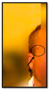
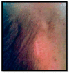
Figure 3
A frame of a picture showing a scarring outcome of a few cm, inveterate, normotrophic, and hypopigmented compared with the surrounding skin. In other frames following Dr. T.A.’s surgery, however, it was possible to observe in the right temporal region, posterior to the eyebrow arch (always used as a repere), a scarring outcome of a few cm, inveterate, normotrophic, hypopigmented compared with the surrounding skin. However, there was no evidence of either the nodular lesion or the pigmented lesion. It was thus possible to infer that the dermatologist had not removed the pigmented lesion in question, but the subcutaneous lesion located superior to it, namely the sebaceous cyst. If in fact, he had removed a melanoma or metastasis of melanoma, having made a small incision, such that a neoplastic lesion (extending into the deeper layers of the skin) could not be completely removed, the melanoma would have reformed in the same location in the immediate following months. Furthermore, since the cyst does not go into regression, it is reasonable to assume that if he had removed the pigmented lesion, there would have been photographic or documentary evidence (in the dermatologic consultation performed during the hospitalization) of a cystic lesion. It is highly likely that the pigmented lesion in question has gone through the phenomenon of regression. In fact, in between 4 and 12 percent of cases, the site of melanoma onset is not recognizable, and the diagnosis is made by the finding of a metastasis in the absence of a clinically evident primary melanoma. This phenomenon can be explained by assuming the presence of a spontaneously regressed cutaneous melanoma. A previous lesion that spontaneously disappeared years before the appearance of metastasis is, in fact, reported in 10–20% of cases in the literature. Histopathologic regression consists, in fact, of partial (segmental) or complete replacement/obliteration of tumor cells associated with mononuclear inflammatory infiltrate, melanophages and/or dermal neovascularization and fibrosis and, therefore, results in the disappearance of the lesion. Photographic images provided by family members together with knowledge regarding the diagnosis of melanoma allowed the case to be reconstructed and the conclusion to be reached that the physician had acted according to science and conscience by removing a benign lesion. The case presented is an example of medico-legal litigation in dermatology [6]. Claims for compensation for failure to diagnose melanoma are very frequent, both because of the inherent characteristics of the neoplasm and because of the physician’s failure to provide detailed documentation. Claims related to cases of omitted diagnosis of melanoma are, in general, due to the alleged failure to diagnose this neoplasm in a timely manner [7,8]. The most common scenarios that result in the misdiagnosis of melanoma are (1) misdiagnosis of nodular melanoma by a dermatologist; (2) misdiagnosis of nodular melanoma by the anatomo-pathologist; (3) incomplete biopsy; (4) melanoma misdiagnosed as “dysplastic nevus with edge involvement”; (5) melanoma misdiagnosed as Spitz’s nevus; (6) unrecognized desmoplastic melanoma and (7) metastatic disease without known primary disease [9]. It will be useful to go over some features of melanoma and its diagnosis in extreme summary. There are different subgroups depending on certain clinical and histopathological features; for the histological classification of melanoma, reference is made to the 2018 WHO classification, which includes, among others, some of the most frequent types of melanoma, including superficial spreading melanoma (SMM), nodular melanoma (NM), lentigo maligna (LM), and acral-lentiginous melanoma (ALM). The clinical diagnosis of melanoma is based on clinical-morphological aspects and dermatoscopic examination [10]. The clinical parameters to be considered are the ABCDE criteria: asymmetry (A), irregularity in borders (B) and color (C), dimensional increase (D), and evolutions within the lesion (E). The ‘dermatoscopic examination can improve diagnostic accuracy by 15–20% over simple clinical observation [11]. Melanoma arises at or contiguous with a preexisting congenital acquired melanocytic nevus, in up to half of the cases [12]. In the remaining cases, it arises in a skin area not the site of melanocytic nevi. The major insidiousness is the occurrence of melanomas at sites other than the skin, such as at the eye or mucosal level (oral cavity, nasal mucosa, vulva and vagina, anus and anorectal canal, penis), or at the nail level. Rare, however, are primary melanomas starting in internal organs [13]. However, the diagnostic confirmation of melanoma is histologic, so in cases of suspected melanoma, a histologic examination is mandated, which allows for the definition of the histotype, the growth phase of the neoplasm, and the evaluation of some parameters that are of fundamental importance with regard to prognosis (Breslow’s thickness, number of mitoses/mmq, ulceration, the possible presence of regression, lymphovascular invasion, and neurotropism) [14,15]. Ranging from 15% to 35%, patients with cutaneous melanoma without metastasis at the time of first diagnosis subsequently develop disease progression. Metastasis can occur by contiguity, by the lymphatic route (to regional lymph nodes or in transit) or by the bloodstream. Spread generally occurs through three modes: direct extension consisting of horizontal and vertical growth stages; lymphatic spread by embolization through intradermal vessels with skin localizations near or distant from the primary site and hematic spread [16]. In 4 to 12 percent of cases, the site of melanoma onset is not recognizable, and the diagnosis is made by the finding of a metastasis, usually a lymph node, in the absence of a clinically evident primary melanoma [8]. This phenomenon, once the presence of mucosal or ocular melanoma is ruled out, can be explained by assuming the presence of a spontaneously regressed cutaneous melanoma or, more rarely, primary lymph node-onset melanoma. A previous lesion that disappeared spontaneously or cauterized or excised several years before the appearance of metastasis is reported in 10–20% of cases [17]. These are the main reasons why dermatology physicians are frequently victims of complaints. Very often, in fact, the diagnosis of melanoma conceals a skin lesion excised a few months earlier, and so the patient or his family members decide to file a complaint against the physician. Very often, the lack of physician protection leads to a shift from clinical prevention activity to litigation prevention. This problem plaguing medicine is not just an Italian problem, but a truly global issue. Even in the United States of America (USA), there have been numerous studies that have addressed the issue of litigation in dermatology. Daniel D. Lydiatt, using the analysis of jury verdicts, conducted an analysis of lawsuits in cutaneous melanoma cases, stating that a failure to diagnose was alleged in 54% of the cases and that, of these, only 48% of the judgments saw a concrete omission of the diagnostic test [7]. This must inevitably give pause to the amount of litigation initiated on unfounded grounds. Unfortunately, so many results in serious harm to the physician, with an inevitable drift of medical work from patient care to personal protection in the view of possible judgment. In patients who present with a diagnosis of metastasis from melanoma in the absence of a primary lesion, following the removal of a ‘benign’ lesion in whom the site of the primary melanoma cannot be located, it may be assumed that the excised or destroyed ‘benign’ lesion was in fact a melanoma [16,18]. One means of avoiding this scenario is to perform a histological examination for all excised lesions, even those that are apparently benign [8]. This will inevitably lead to increased costs and longer waiting lists for histologic diagnosis, with the downside that in those truly affected by neoplastic disease there will be delayed initiation of therapy. However, is this really the direction to go? Is it right to put judicial protection first rather than patient care? In this scenario of “malpractice hunting,” the physician must be better protected and must, therefore, have the tools to defend himself, considering that the diagnosis of melanoma, as we have seen in our case, must be timely and the survival rate depends precisely on the celerity of the diagnosis [19,20,21,22]. Actually, it would suffice to comply with a protocol to be followed in the case of skin lesions that need removal. The physician in charge must create a file for each patient in which, in addition to noting the medical history, he or she must include a detailed description of the lesion to be removed and, with the patient’s consent, attach photographic documentation of it. For each lesion removed, it will be helpful to describe: the type, shape, color, size, and anatomic location, indicate whether it is a lesion detected in relation to the surrounding skin, and approximate date of the appearance of the lesion. It will, in addition, be useful to take photographs in which the location of the lesion can be pinpointed, and its macroscopic features identified [17]. Even more useful would be to also attach any photographs taken with the use of a dermatoscope. Such a simple stratagem would make it possible not to overshadow patient care, while also ensuring elements of high evidentiary power in court. The possibility of attesting to what has been conducted with documentary elements (photos and description), guarantees protection for the physician who will, therefore, be able to always operate in the best interest of the patient, but in full self-defense in the possible judicial context. If in the case presented, the dermatology physician had operated with these indications, the doubt would probably not have arisen that he had removed a neoplastic lesion. The case was, in fact, able to be resolved occasionally with the help of amateur photographs of the deceased. So much demonstrates the importance of iconographic documentation, especially when excising benign lesions. Small shrewdness could, therefore, secure the physician, and avoid numerous claims, attested, however, histological examination is mandated always since clinical and dermoscopic features are never diagnostic, but only indicative, sometimes with excellent reliability, but in other cases, precisely because of the intrinsic characteristics of some skin lesions, absolutely unreliable.


