Review
. 2023 Mar 16;13(6):1127.
doi: 10.3390/diagnostics13061127.

Hydatid Disease: A Radiological Pictorial Review of a Great Neoplasms Mimicker

Affiliations
Review

Hydatid Disease: A Radiological Pictorial Review of a Great Neoplasms Mimicker

Sultan Abdulwadoud Alshoabi et al. Diagnostics (Basel). .

Abstract

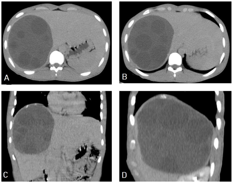
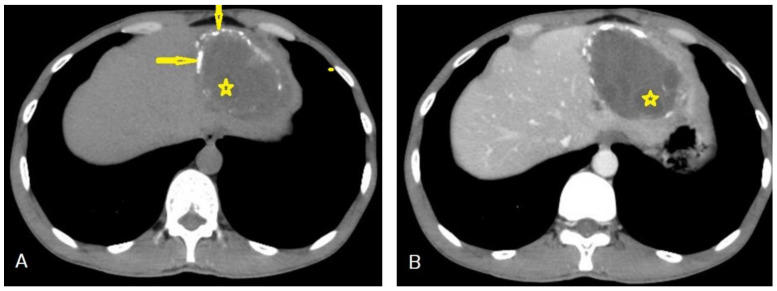
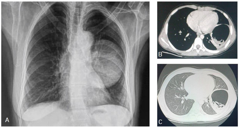
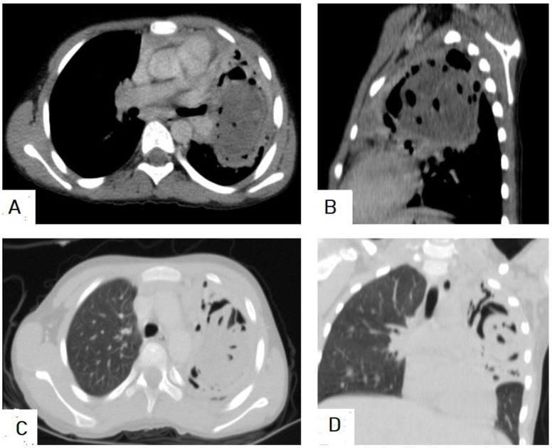
Hydatid cyst is a common name for the larval stage of a tapeworm species of Echinococcus granulosus, which is transmitted from animals to humans via the fecal-oral route. Hydatid cysts predominantly affect the liver (75%), followed by the lung (15%), and they can affect many organs in the human body. Medical imaging modalities are the keystone for the diagnosis of hydatid cysts with high sensitivity and specificity. Ultrasound imaging with high resolution is the first choice for diagnosis, differential diagnosis, staging, establishing a role in interventional management, and follow-up, and it can differentiate Type I hydatid cysts from simple liver cysts. Unenhanced computed tomography (CT) is indicated where or when an ultrasound is unsatisfactory, such as with chest or brain hydatid cysts, when detecting calcification, and in obese patients. Magnetic resonance imaging (MRI) is superior for demonstrating cyst wall defects, biliary communication, neural involvement, and differentiating hydatid cysts from simple cysts using diffusion-weighted imaging (DWI) sequences. According to the phase of growth, hydatid cysts occur in different sizes and shapes, which may mimic benign or malignant neoplasms and may create diagnostic challenges in some cases. Hydatid cysts can mimic simple cysts, choledochal cysts, Caroli's disease, or mesenchymal hamartomas of the liver. They can mimic lung cystic lesions, mycetoma, blood clots, Rasmussen aneurysms, and even lung carcinomas. Differential diagnosis can be difficult for arachnoid cysts, porencephalic cysts, pyogenic abscesses, and even cystic tumors of the brain, and can create diagnostic dilemmas in the musculoskeletal system.

Keywords: calcified cyst; cyst with daughter cysts; cyst with floating membranes; cyst with waterlily sign; larval stage of Echinococcus granulosus; spoke-wheel appearance; unilocular simple cyst.


Conflict of interest statement

The authors declare no conflict of interest.

Figures

Figure 1
Figure 1
Method of human infection with Echinococcus granulosus.
Figure 2
Figure 2
Images of gross hydatid cysts from outside (A), and inside (B) of two different cysts.
Figure 3
Figure 3
Contrast-enhanced computed tomography of a 15-year-old female presenting with abdominal pain showing a well-circumscribed, unilocular, non-enhanced cystic lesion in the right lobe of the liver with typical “double-wall sign” of hydatid cyst (arrows).
Figure 4
Figure 4
Ultrasound images of a 35-year-old female showing a well-circumscribed, unilocular, anechoic cystic lesion measuring 10 cm with no internal septation or solid component centered in segment IV of the right hepatic lobe, exerting slight compression on the gallbladder with no remarkable biliary dilatation or frank calcifications. Picture is in keeping with hydatid cyst stage CE1 (WHO classification).
Figure 5
Figure 5
Ultrasound image of a 17-year-old female presenting with upper abdominal pain shows an 11 × 6 cm well-demarcated, mild lobulated margin with multiple internal septations of higher attenuation seen in segment VI of the right lower hepatic lobe, descended in front, lateral to the right kidney, and exerting mild kidney indentation. Picture is in keeping with the hydatid cyst CE2 (WHO classification).
Figure 6
Figure 6
Ultrasound images of the liver of a 10-year-old male presenting with abdominal pain showing 15 × 12 cm well-circumscribed, cystic lesion thin echogenic wall replacing the V, VI, and VII segments of right hepatic lobe containing multiple daughter cysts and echogenic matrices (A,B), which are typical images of hydatid cysts in stage CE3b (WHO classification). The daughter cysts appeared more obviously on the high-resolution images of the high-frequency linear transducer (C,D).
Figure 7
Figure 7
Selected CT images of a 35-year-old female showing a well-circumscribed, round, unilocular hypodense cystic lesion centered in segment IV of the right hepatic lobe, measuring 10 × 7.7 × 6.3 cm with no internal septation or solid component, no frank calcifications, or enhanced internal component after contrast administration, which is a typical image of hydatid cyst Type I.
Figure 8
Figure 8
Selected axial (A) and coronal (B) CT images of a 60-year-old female showing multiple well-defined cystic lesions occupying the liver: about five lesions in the right lobe, where the largest one measured 17 × 9.2 × 15.3 cm (long arrows), occupying the right posterior lobe, and two cysts occupying the left lobe, where the largest one measured 11.4 × 10.3 × 8.7 cm (short arrows). Most of the lesions contain multiple vesicular cysts inside them (daughter cysts). The lesions exert a compression effect on the portal vein, stomach, and ascending colon, intending to reach the right kidney upper pole. This is a typical picture of an active-stage hydatid cyst Type IIA.
Figure 9
Figure 9
Selected axial (A,B) and coronal (C,D) CT images of a 16-year-old male presenting with abdominal pain and distension showing a well-defined cystic lesion in the right lobe of the liver, containing multiple vesicular cysts (daughter cysts) and hyperdense matrix, and forming the “spoke-wheel” appearance of a hydatid cyst.
Figure 10
Figure 10
Selected axial (A) and sagittal (B) CT image of a 10-year-old male presenting with abdominal pain shows an enlarged liver with a 15 × 12 cm thin-wall cystic lesion seen replacing the V, VI, and VII segments of right hepatic lobe containing multiple daughter cysts, which is typical picture of a hydatid cyst with normal biliary tree and portal vein. Another similar thin-wall cystic lesion measuring 5.5 × 3.7 cm contains multiple daughter cysts, which is a typical picture of hydatid cysts of the spleen. Additionally, showing multiple similar cystic lesions measuring 11 × 16 cm in the right upper abdominal cavity causing mass effect in the displacement of the right kidney, and another three smaller cystic lesions in the right subphrenic space measuring 3.8 × 5.2 cm, 4 × 2 cm, and 4 × 1.5 cm. Moreover, showing another similar cystic lesion at the recto-vesical pouch measuring about 6 × 4 cm. The above findings represent a typical case of liver, spleen, and peritoneal hydatidosis and Type IIB.
Figure 11
Figure 11
Selected axial non-enhanced (A), and contrast-enhanced (B) CT images of the abdomen of an adult male patient showing a well-circumscribed hypodense cystic lesion with multiple daughter cysts inside it (stars), with stippled calcification of the wall (arrow), and with no enhancement after contrast administration. Picture of hydatid cyst Type IIC.
Figure 12
Figure 12
Selected axial images of abdominal CT of a 60-year-old female showing a well-circumscribed unilocular cystic lesion in the right kidney with calcified margins (arrows), no mass effect, and no daughter cysts inside it, which is typical of hydatid cyst Type III.
Figure 13
Figure 13
Selected axial (A) and sagittal (B) CT images of a 50-year-old female presenting with right upper quadrant pain, showing a well-circumscribed unilocular cystic lesion in the right lobe of the liver with calcified margins and no daughter cysts inside it, which is typical of inactive hydatid cyst Type III.
Figure 14
Figure 14
Selected CT images of a 26-year-old male showing multiple well-defined grouped cysts in the right lobe of the liver, the largest about 4 cm, seen on axial (A) section, with partial peripheral calcification seen on axial (B) section. There are multiple irregular, ill-defined small cysts around the confluence of the main hepatic bile ducts seen on axial (C) section with moderate dilatation of the common bile duct and mild dilatation of the intrahepatic biliary tree seen on coronal (D) section. Picture is in keeping with biliary tree seeding from the liver hydatid cyst.
Figure 15
Figure 15
Selected CT images of a 40-year-old male presenting with cough and dyspnea showing a ruptured liver hydatid cyst complicated with disastrous dissemination into the peritoneal cavity seen on the axial section (A), and bilateral chest seeding seen on axial (B), sagittal (C), and coronal (D) sections. Picture is in keeping with peritoneal and bilateral chest seeding from the liver hydatid cyst.
Figure 16
Figure 16
Selected CT images of an 80-year-old man presenting with chest pain, showing about 6.8 × 4 cm multiloculated and peripherally enhanced collection and seeming to arise from the subcapsular segment-VII of the right lobe of liver with disrupted capsule and exophytic tracking component to subphrenic region into the posterior perinephric and pararenal spaces seen on axial (A,B) sections, and extending across the proximal psoas muscle for 7.5 cm caudally seen on sagittal (C) and coronal (D) sections. Superiorly, the lesion tracks the right hemidiaphragm into the lung, where there is a well-circumscribed mass-like lesion measured at 8.7 × 6.3 × 5 cm with thickened wall with obvious bronchovascular markings seen extended within the lesion in the adjacent lower lobe area seen on sagittal (C), coronal (D), axial (E), and sagittal lung window (F) sections. The picture suggests ruptured hepatic hydatid cyst tracking to the retroperitoneaum, psoas muscle, and lung, with superimposed psoas abscess formation.
Figure 17
Figure 17
Selected CT images of a 60-year-old male with renal impairment presenting with abdominal pain and distention, showing 8.6 × 8.2 cm hydatid cyst with multiple daughter cysts in the lower lobe of the right lung, indenting the right hemidiaphragm, and compressing the lower liver seen on axial (A) and coronal (D) sections. Additionally, showing multiple abdominal and pelvic large hydatid cysts with multiple internal daughter cysts compressing bilateral ureters, causing moderate bilateral hydronephrosis, and compressing the urinary bladder anteriorly seen on axial (B,C), coronal (D,E), and sagittal (F) sections. Pictures from Type IIA with typical “spoke-wheel appearance” occurring on images (AD).
Figure 18
Figure 18
Selected CT images of a 13-year-old patient presenting with abdominal distention and feeling mass showing a 16 × 15 × 11 cm well-circumscribed, multiloculated cystic lesion in the right side of abdominal and upper pelvic cavities as seen in axial (A,B) sections, exerting mass effect in the form of bilateral mild hydronephrosis, displacing the bowel loops laterally, and compressing the urinary bladder as seen in the sagittal (C) and coronal (D) sections. The cyst had a narrow neck-like (arrow) at the superolateral aspect connected with other cystic lesions in segment-VI of the liver. The above findings are in keeping with a large abdominal hydatid cyst.
Figure 19
Figure 19
Selected axial sections of CT images of a 30-year-old female with a previous surgery for a liver hydatid cyst presenting with abdominal pain showing remnants of the hydatid cyst (arrows) and another hydatid cyst (arrow heads).
Figure 20
Figure 20
Chest X-ray of a 9-year-old male patient showing a well-defined oval shape with smooth margins and a large mass in the left lung (A). Selected axial CT images, mediastinal window (B), and lung window (C) show a well-circumscribed smooth margin intrapulmonary mass measuring 8.4 × 6.4 cm with fluid and gas contents with twisted-linear structures floating within the fluid contents of the cyst, forming a “water lily sign”, and representing hydatid cyst with detachment of the germinal membrane of the endocyst. The mass causes a mass effect in the form of compression of the lower lobe of the left lung.
Figure 21
Figure 21
Selected sections of chest CT images of a 10-year-old female presenting with cough, dyspnea, and hemoptysis showing a hypodense lesion in the left lung with thick-enhancing wall seen on axial (A) and sagittal (B) mediastinal window sections. The “air-bubble” sign inside the lesion is seen in the mediastinal window sections (A,B), and on the lung window axial (C) and coronal (D) sections. Picture is in keeping with ruptured hydatid cyst.
Figure 22
Figure 22
Selected axial (A) and coronal (B) CT images of a 25-year-old male with left upper abdominal pain and distention, showing a 15 × 13 cm well-circumscribed, unilocular cystic lesion in the spleen that causes a mass effect in the form of compression and downward displacement of the left kidney (arrows). The lesion showed homogeneous contents, no septations, no calcification, and no enhancement after contrast administration, which is a typical picture of a hydatid cyst of the spleen.
Figure 23
Figure 23
Selected CT images of a 12-year-old male presenting with signs of increased intracranial pressure, showing a large well-circumscribed cyst in the right cerebral hemisphere that caused a mass effect in the form of compression of the ipsilateral lateral ventricle complicating with ipsilateral hydrocephalus. The cyst showed no surrounding edema, no calcification (A), and no enhancement after contrast administration (B), which is a typical feature of cerebral hydatid cysts.
Figure 24
Figure 24
Selected images of thigh MRI of a 23-year-old male showing an 11 × 8 × 8 cm heterogeneous complex cystic lesion within the anteromedial intramuscular compartment of the upper right thigh with thickened edematous adjacent subcutaneous fat containing multiple small daughter cysts appearing hypointense on T1-weighted images (A), hyperintense on T2-weighted images (B), hyperintense on fat-suppression sequences (C), and marginal wall enhancement on T1 with contrast (D), which is typical appearance of intramuscular hydatid cyst with superimposed infection and impending rapture.
Figure 25
Figure 25
Selected images of pelvic MRI of a 75-year-old female presenting with pelvic pain, difficulty defecating, and micturition, which shows a 12 × 9 cm well-circumscribed, multiloculated cystic lesion in the left presacral and paranal, with ischiorectal fossa causing mass effect in the form of displacing the anal canal and rectum to the right side. The lesion appears hypointense on axial and sagittal T1-weighted images (A,C), and hyperintense on axial and sagittal T2-weighted images (B,D), with multiple cysts inside it, which is a typical picture of an intramuscular hydatid cyst.

References

    1. Agudelo Higuita N.I., Brunetti E., McCloskey C. Cystic Echinococcosis. J. Clin. Microbiol. 2016;54:518–523. doi: 10.1128/JCM.02420-15. - DOI - PMC - PubMed
    1. Zhang T., Li B., Liu Y., Liu S. Risk Factors Associated With Echinococcosis in the General Chinese Population: A Meta-Analysis and Systematic Review. Front. Public Health. 2022;10:821265. doi: 10.3389/fpubh.2022.821265. - DOI - PMC - PubMed
    1. Lodhia J., Chugulu S., Sadiq A., Msuya D., Mremi A. Giant isolated hydatid lung cyst: Two case reports. J. Med. Case Rep. 2020;14:200. doi: 10.1186/s13256-020-02524-4. - DOI - PMC - PubMed
    1. Torgerson P.R., Devleesschauwer B., Praet N., Speybroeck N., Willingham A.L., Kasuga F., Rokni M.B., Zhou X.N., Fèvre E.M., Sripa B., et al. World Health Organization Estimates of the Global and Regional Disease Burden of 11 Foodborne Parasitic Diseases, 2010: A Data Synthesis. PLoS Med. 2015;12:e1001920. doi: 10.1371/journal.pmed.1001920. - DOI - PMC - PubMed
    1. Da Silva A.M. Human echinococcosis: A neglected disease. Gastroenterol. Res. Pract. 2010;2010:583297. doi: 10.1155/2010/583297. - DOI - PMC - PubMed

LinkOut - more resources