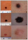
Is Pediatric Melanoma Really That Different from Adult Melanoma? A Multicenter Epidemiological, Clinical and Dermoscopic Study
- PMID: 36980721
- PMCID: PMC10046848
- DOI: 10.3390/cancers15061835
Is Pediatric Melanoma Really That Different from Adult Melanoma? A Multicenter Epidemiological, Clinical and Dermoscopic Study
Abstract
Purpose: To improve the diagnostic accuracy and optimal management of pediatric melanomas.
Methods: We conducted a retrospective descriptive, multicenter study of the epidemiological, clinical, and dermoscopic characteristics of histopathologically proven melanomas diagnosed in patients less than 18 years old. Data on sociodemographic variables, clinical and dermoscopic characteristics, histopathology, local extension, therapy and follow-up, lymph node staging, and outcome were collected from the databases of three Italian dermatology units. We performed a clinical evaluation of the morphological characteristics of each assessed melanoma, using both classic ABCDE criteria and the modified ABCDE algorithm for pediatric melanoma to evaluate which of the two algorithms best suited our series.
Results: The study population consisted of 39 patients with a histologically confirmed diagnosis of pediatric melanoma. Comparing classic ABCDE criteria with the modified ABCDE algorithm for pediatric melanomas, the modified pediatric ABCDE algorithm was less sensitive than the conventional criteria. Dermoscopically, the most frequent finding was the presence of irregular streaks/pseudopods (74.4%). When evaluating the total number of different suspicious dermoscopy criteria per lesion, 64.1% of the lesion assessments recognized two dermoscopic characteristics, 20.5% identified three, and 15.4% documented four or more assessments.
Conclusions: Contrary to what has always been described in the literature, from a clinical point of view, about 95% of our cases presented in a pigmented and non-amelanotic form, and these data must be underlined in the various prevention campaigns where pediatric melanoma is currently associated with a more frequently amelanotic form. All the pediatric melanomas analyzed presented at least two dermoscopic criteria of melanoma, suggesting that this could be a key for the dermoscopic diagnosis of suspected pediatric melanoma, making it possible to reach an early diagnosis even in this age group.
Keywords: children; dermoscopy; estrogen; melanoma; skin cancer.
Conflict of interest statement
The authors declare no conflict of interest.
Figures


References
-
- Garbe C., Amaral T., Peris K., Hauschild A., Arenberger P., Bastholt L., Fargnoli M.C., Grob J.J., Höller C., Kaufmann R., et al. European Dermatology Forum (EDF), the European Association of Dermato-Oncology (EADO), and the European Organization for Research and Treatment of Cancer (EORTC). European consensus-based interdisciplinary guideline for melanoma. Part 1: Diagnostics—Update 2019. Eur. J. Cancer. 2020;126:141–158. doi: 10.1016/j.ejca.2019.11.014. - DOI - PubMed
LinkOut - more resources
Full Text Sources

