

Zeb2 DNA-Binding Sites in Neuroprogenitor Cells Reveal Autoregulation and Affirm Neurodevelopmental Defects, Including in Mowat-Wilson Syndrome
- PMID: 36980900
- PMCID: PMC10048071
- DOI: 10.3390/genes14030629
Zeb2 DNA-Binding Sites in Neuroprogenitor Cells Reveal Autoregulation and Affirm Neurodevelopmental Defects, Including in Mowat-Wilson Syndrome
Abstract
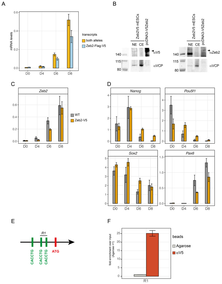
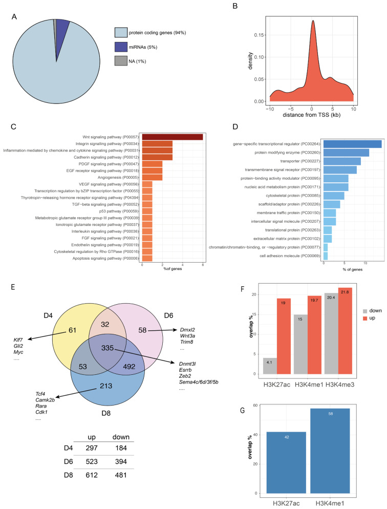
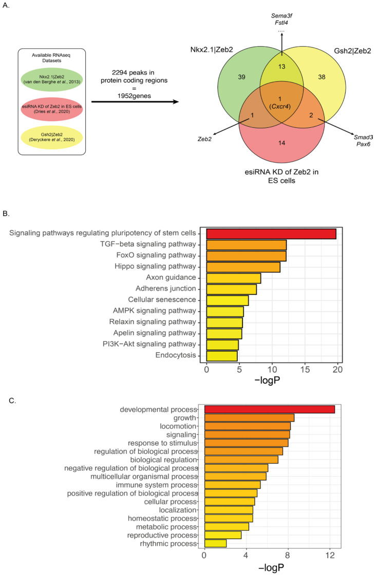
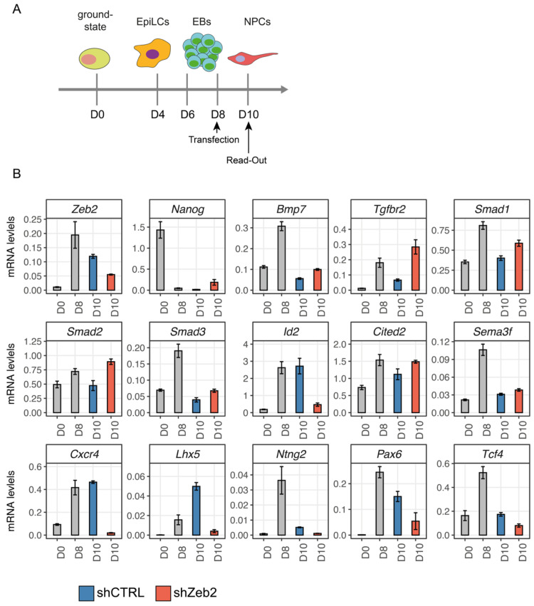
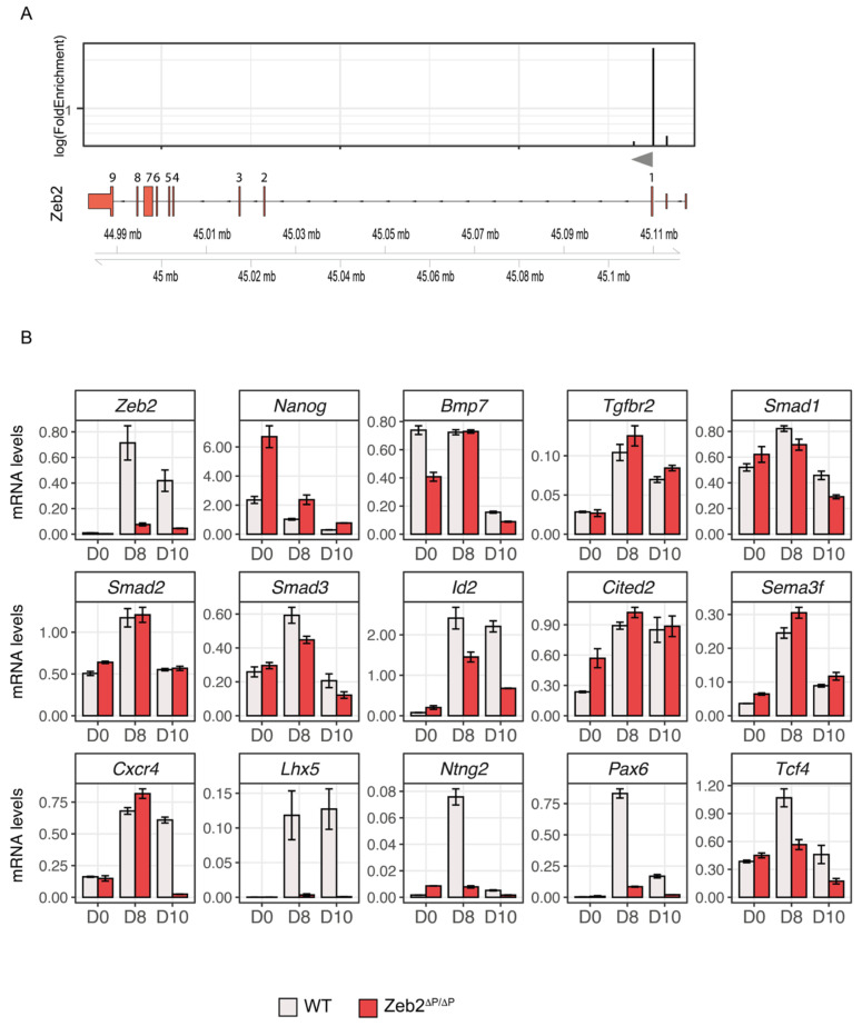
Functional perturbation and action mechanism studies have shown that the transcription factor Zeb2 controls cell fate decisions, differentiation, and/or maturation in multiple cell lineages in embryos and after birth. In cultured embryonic stem cells (ESCs), Zeb2's mRNA/protein upregulation is necessary for the exit from primed pluripotency and for entering general and neural differentiation. We edited mouse ESCs to produce Flag-V5 epitope-tagged Zeb2 protein from one endogenous allele. Using chromatin immunoprecipitation coupled with sequencing (ChIP-seq), we mapped 2432 DNA-binding sites for this tagged Zeb2 in ESC-derived neuroprogenitor cells (NPCs). A new, major binding site maps promoter-proximal to Zeb2 itself. The homozygous deletion of this site demonstrates that autoregulation of Zeb2 is necessary to elicit the appropriate Zeb2-dependent effects in ESC-to-NPC differentiation. We have also cross-referenced all the mapped Zeb2 binding sites with previously obtained transcriptome data from Zeb2 perturbations in ESC-derived NPCs, GABAergic interneurons from the ventral forebrain of mouse embryos, and stem/progenitor cells from the post-natal ventricular-subventricular zone (V-SVZ) in mouse forebrain, respectively. Despite the different characteristics of each of these neurogenic systems, we found interesting target gene overlaps. In addition, our study also contributes to explaining developmental disorders, including Mowat-Wilson syndrome caused by ZEB2 deficiency, and also other monogenic syndromes.
Keywords: Mowat-Wilson syndrome; Zeb2; chromatin immunoprecipitation sequencing; embryonic stem cells; neural differentiation; neurodevelopmental disorder; syndromes; target genes; transcription factor; transcriptomics.
Conflict of interest statement
The authors declare no conflict of interest.
Figures
Similar articles
-
ZEB2, the Mowat-Wilson Syndrome Transcription Factor: Confirmations, Novel Functions, and Continuing Surprises.Genes (Basel). 2021 Jul 3;12(7):1037. doi: 10.3390/genes12071037. Genes (Basel). 2021. PMID: 34356053 Free PMC article. Review.
-
Targeted chromatin conformation analysis identifies novel distal neural enhancers of ZEB2 in pluripotent stem cell differentiation.Hum Mol Genet. 2020 Aug 29;29(15):2535-2550. doi: 10.1093/hmg/ddaa141. Hum Mol Genet. 2020. PMID: 32628253 Free PMC article.
-
Zeb2 Regulates Cell Fate at the Exit from Epiblast State in Mouse Embryonic Stem Cells.Stem Cells. 2017 Mar;35(3):611-625. doi: 10.1002/stem.2521. Epub 2016 Nov 8. Stem Cells. 2017. PMID: 27739137 Free PMC article.
-
Requirement of the Mowat-Wilson Syndrome Gene Zeb2 in the Differentiation and Maintenance of Non-photoreceptor Cell Types During Retinal Development.Mol Neurobiol. 2019 Mar;56(3):1719-1736. doi: 10.1007/s12035-018-1186-6. Epub 2018 Jun 19. Mol Neurobiol. 2019. PMID: 29922981
-
Role of Zeb2/Sip1 in neuronal development.Brain Res. 2019 Feb 15;1705:24-31. doi: 10.1016/j.brainres.2018.09.034. Epub 2018 Sep 25. Brain Res. 2019. PMID: 30266271 Review.
Cited by
-
Editorial for the Molecular Mechanisms in Neurodevelopmental Disorders Special Issue.Genes (Basel). 2023 Sep 4;14(9):1762. doi: 10.3390/genes14091762. Genes (Basel). 2023. PMID: 37761902 Free PMC article.
-
The Multifaceted Roles of Zinc Finger Proteins in Pluripotency and Reprogramming.Int J Mol Sci. 2025 May 26;26(11):5106. doi: 10.3390/ijms26115106. Int J Mol Sci. 2025. PMID: 40507915 Free PMC article. Review.
-
Identification of the DNA methylation signature of Mowat-Wilson syndrome.Eur J Hum Genet. 2024 Jun;32(6):619-629. doi: 10.1038/s41431-024-01548-4. Epub 2024 Feb 13. Eur J Hum Genet. 2024. PMID: 38351292 Free PMC article.
References
-
- Remacle J.E., Kraft H., Lerchner W., Wuytens G., Collart C., Verschueren K., Smith J.C., Huylebroeck D. New mode of DNA binding of multi-zinc finger transcription factors: DeltaEF1 family members bind with two hands to two target sites. EMBO J. 1999;18:5073–5084. doi: 10.1093/emboj/18.18.5073. - DOI - PMC - PubMed
-
- Verschueren K., Remacle J.E., Collart C., Kraft H., Baker B.S., Tylzanowski P., Nelles L., Wuytens G., Su M.T., Bodmer R., et al. SIP1, a novel zinc finger/homeodomain repressor, interacts with Smad proteins and binds to 5’-CACCT sequences in candidate target genes. J. Biol. Chem. 1999;274:20489, 20498. doi: 10.1074/jbc.274.29.20489. - DOI - PubMed
Publication types
MeSH terms
Substances
Supplementary concepts
LinkOut - more resources
Full Text Sources
Research Materials
