Review
. 2023 Mar 8;24(6):5155.
doi: 10.3390/ijms24065155.

Intracoronary Imaging of Coronary Atherosclerotic Plaque: From Assessment of Pathophysiological Mechanisms to Therapeutic Implication

Affiliations
Review

Intracoronary Imaging of Coronary Atherosclerotic Plaque: From Assessment of Pathophysiological Mechanisms to Therapeutic Implication

Filippo Luca Gurgoglione et al. Int J Mol Sci. .

Abstract

Atherosclerotic cardiovascular disease is the leading cause of morbidity and mortality worldwide. Several cardiovascular risk factors are implicated in atherosclerotic plaque promotion and progression and are responsible for the clinical manifestations of coronary artery disease (CAD), ranging from chronic to acute coronary syndromes and sudden coronary death. The advent of intravascular imaging (IVI), including intravascular ultrasound, optical coherence tomography and near-infrared diffuse reflectance spectroscopy has significantly improved the comprehension of CAD pathophysiology and has strengthened the prognostic relevance of coronary plaque morphology assessment. Indeed, several atherosclerotic plaque phenotype and mechanisms of plaque destabilization have been recognized with different natural history and prognosis. Finally, IVI demonstrated benefits of secondary prevention therapies, such as lipid-lowering and anti-inflammatory agents. The purpose of this review is to shed light on the principles and properties of available IVI modalities along with their prognostic significance.

Keywords: biological mechanisms; coronary artery disease; intracoronary imaging; plaque healing; plaque vulnerability; secondary prevention therapies.


Conflict of interest statement

The authors declare no conflict of interest.

Figures

Figure 1
Figure 1
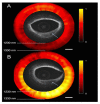
IVUS images of coronary plaque phenotype. (A) Example of a calcific plaque, depicted as a bright leading circumferential structure ((A), white arrow) with deeper shadowing ((A), white star). (B) Example of predominantly fibro-fatty plaque, depicted as a structure showing less echogenicity than the surrounding adventitia with a spotty calcification ((B), white arrow), characterized by a focal hyperechoic signal and deeper shadowing.
Figure 2
Figure 2
Examples of IVUS-based mechanisms of plaque destabilization. (A) Example of a calcific nodule, depicted as a convex-shape protrusion into the vessel lumen (5 to 7 o’clock positions) with hyperechoic appearance ((A), white arrow) with deeper shadowing ((A), white star). (B) Example of a suspected ruptured plaque in a patient with ACS, labelled as a cavity that communicates with the vessel lumen with a tear in the fibrous cap ((B), white star).
Figure 3
Figure 3
OCT images of coronary plaque phenotype. (A) Pathological intimal thickening, depicted as a homogeneous signal-rich thick intimal band composed of fibrous tissue. (B) Calcific plaque ((B), white arrow), depicted as a low-backscattering structure, as compared to surrounding adventitia, with sharply delineated borders. (C) TCFA, depicted as a low-density structure with diffuse borders covered by a thin fibrous cap. Macrophages ((C), white arrow), identified as signal-rich distinct or confluent punctate regions that exceed the intensity of background speckle noise, are often found in TCFAs.
Figure 4
Figure 4
Examples of OCT-derived mechanisms of plaque destabilization. (A) Example of a ruptured plaque, characterized by the evidence of a cavity with a clear discontinuity of the fibrous cap ((A), white arrow). (B) Example of a definite eroded plaque, characterized by a luminal thrombus ((B), white arrow) overlying a plaque without evidence of fibrous cap disruption. (C) Example of a calcific nodule ((C), white arrow), depicted as single of multiple regions of calcium that protrude into the lumen with fibrous cap disruption.
Figure 5
Figure 5
An example of OCT-derived healed plaque defined as a plaque presenting with one or more signal-rich layers (6 to 11 o’clock position, white arrow) of different optical density and a clear demarcation from underlying components in at least three consecutive frames along the entire plaque.
Figure 6
Figure 6
Illustration of the NIRS chemogram and block chemogram. The x-axis of the chemogram represents the spatial location along the long axis of the vessel. The y-axis of the chemogram represents circumferential position. Red and yellow regions correspond to coronary segments with, respectively, a low and high probability of LRP. Reused with permission from AME Publishing Company [80].
Figure 7
Figure 7
Selection of a region of interest (50 mm segment of the targeted artery, white lines) and the quantification of maxLCBI4mm (the 4 mm segment within the region of interest containing the greatest LCBI, blue lines) by the NIRS software. Reused with permission from AME Publishing Company [80].
Figure 8
Figure 8
OCT-NIRS images of cadaver coronary artery ex vivo. (A) A lesion with low lipid content, characterized by OCT-low backscattering and NIRS-low signal. (B) A lipid-rick lesion, characterized by OCT-low backscattering and NIRS-high signal. Reprinted with permission from ref. [97] © The Optical Society.
Figure 9
Figure 9
Graphical summary of clinical applications of intracoronary imaging.

References

    1. Roth G.A., Mensah G.A., Johnson C.O., Addolorato G., Ammirati E., Baddour L.M., Barengo N.C., Beaton A.Z., Benjamin E.J., Benziger C.P., et al. GBD-NHLBI-JACC Global Burden of Cardiovascular Diseases Writing Group. Global Burden of Cardiovascular Diseases and Risk Factors, 1990–2019: Update From the GBD 2019 Study. J. Am. Coll. Cardiol. 2020;76:2982–3021. doi: 10.1016/j.jacc.2020.11.010. Erratum in J. Am. Coll. Cardiol. 2021, 77, 1958–1959. - DOI - PMC - PubMed
    1. Otsuka F., Yasuda S., Noguchi T., Ishibashi-Ueda H. Pathology of coronary atherosclerosis and thrombosis. Cardiovasc. Diagn. Ther. 2016;6:396–408. doi: 10.21037/cdt.2016.06.01. - DOI - PMC - PubMed
    1. Frąk W., Wojtasińska A., Lisińska W., Młynarska E., Franczyk B., Rysz J. Pathophysiology of Cardiovascular Diseases: New Insights into Molecular Mechanisms of Atherosclerosis, Arterial Hypertension, and Coronary Artery Disease. Biomedicines. 2022;10:1938. doi: 10.3390/biomedicines10081938. - DOI - PMC - PubMed
    1. Knuuti J., Wijns W., Saraste A., Capodanno D., Barbato E., Funck-Brentano C., Prescott E., Storey R.F., Deaton C., Cuisset T., et al. 2019 ESC Guidelines for the diagnosis and management of chronic coronary syndromes. Eur. Heart J. 2020;41:407–477. doi: 10.1093/eurheartj/ehz425. Erratum in Eur. Heart J. 2020, 41, 4242. - DOI - PubMed
    1. Collet J.-P., Thiele H., Barbato E., Barthélémy O., Bauersachs J., Bhatt D.L., Dendale P., Dorobantu M., Edvardsen T., Folliguet T., et al. 2020 ESC Guidelines for the management of acute coronary syndromes in patients presenting without persistent ST-segment elevation. Eur. Heart J. 2021;42:1289–1367. doi: 10.1093/eurheartj/ehaa575. Erratum in Eur. Heart J. 2021, 42, 1908; Erratum in Eur. Heart J. 2021, 42, 1925; Erratum in Eur. Heart J. 2021. - DOI - PubMed

MeSH terms