Review
. 2023 Mar 8;24(6):5182.
doi: 10.3390/ijms24065182.

Sensing and Stimulation Applications of Carbon Nanomaterials in Implantable Brain-Computer Interface

Affiliations
Review

Sensing and Stimulation Applications of Carbon Nanomaterials in Implantable Brain-Computer Interface

Jinning Li et al. Int J Mol Sci. .

Abstract

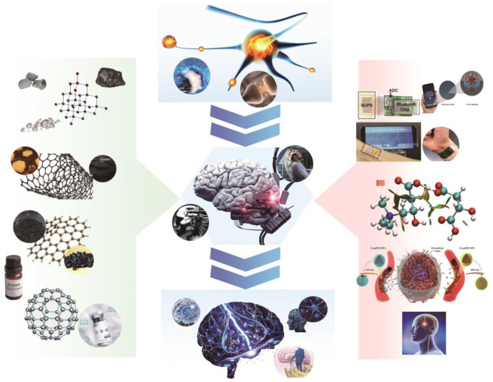
Implantable brain-computer interfaces (BCIs) are crucial tools for translating basic neuroscience concepts into clinical disease diagnosis and therapy. Among the various components of the technological chain that increases the sensing and stimulation functions of implanted BCI, the interface materials play a critical role. Carbon nanomaterials, with their superior electrical, structural, chemical, and biological capabilities, have become increasingly popular in this field. They have contributed significantly to advancing BCIs by improving the sensor signal quality of electrical and chemical signals, enhancing the impedance and stability of stimulating electrodes, and precisely modulating neural function or inhibiting inflammatory responses through drug release. This comprehensive review provides an overview of carbon nanomaterials' contributions to the field of BCI and discusses their potential applications. The topic is broadened to include the use of such materials in the field of bioelectronic interfaces, as well as the potential challenges that may arise in future implantable BCI research and development. By exploring these issues, this review aims to provide insight into the exciting developments and opportunities that lie ahead in this rapidly evolving field.

Keywords: carbon nanomaterials; implantable brain–computer interface; sensing and stimulation.


Conflict of interest statement

The authors declare no conflict of interest.

Figures

Figure 2
Figure 2
(a) SEM image of CNT−coated MEA electrode (∼20 µm diameter). The crater was formed by ablating the overlying dielectric layer to access the indium−tin oxide conductor. Inset: high magnification reveals the porous character of the CNT coating. (b,c) The CNT coating led to a 23−fold decrease in impedance and a 45−fold increase in charge transfer. Reprinted with permission from [40] Copyright © 2023, Springer Nature. (d) MWNT coating thickness as a function of deposition parameters. (e,f) Comparison of electrochemical properties on a per unit film thickness basis. The coating−thickness normalized measurements were obtained from electrochemical impedance spectroscopy and cyclic voltammetry. Reprinted with permission from [70] Copyright © 2023 American Chemical Society. (g) The graphene/Ag electrodes in different scale magnifications. (h,i) Bode plot of (h) impedance and (i) phase of electrodes as a function of the frequency. (j) diamond−coated CFs. Reprinted with permission from [78] Copyright © 2023 American Chemical Society. (k) Optical image of a single diamondecoated CF electrode encapsulated in a glass pipette with the SEM image of the electrode tip shown in (j) inset. (l) Specific capacitance increased from 0.263 ± 0.168 mF/cm2 on CF to 20.90 ± 10.30 mF/cm2 on D−CF electrodes; CIC of D−CF was 238 times larger than that of CF, which increased from 0.105 ± 0.067 mC/cm2 to 25.08 ± 12.37 mC/cm2. Reprinted with permission from [81] Copyright © 2023 Elsevier.
Figure 3
Figure 3
(a) Schematic illustration of a flexible graphene neural electrode array. (b) EIS results for 50 × 50 µm2 Au, G, and doped graphene samples. Measurement results are shown with symbols, and regression results are shown with solid lines for impedance magnitude (top figure) and phase (bottom figure) plots. The impedance magnitude (top figure) significantly decreased with the doping of graphene, more prominently for frequencies lower than 1 kHz. Reprinted with permission from [75] Copyright © 2023, Springer Nature. (c) Schematic drawing of ERG recording with the GRACE device. (d) Photographs of a GRACE device made from G−quartz. Scale bar, 3 mm. The image in the inset demonstrates the high softness of the GRACE device. Reprinted with permission from [77] Copyright © 2023, Springer Nature. (e) Porous graphene is fabricated with laser pyrolysis. (f) A picture of the 64−channel porous graphene electrodes array. The scale bar: 1 cm. (g) A resistive flex sensor spanning the knee joint. The inset illustrates the flexibility of the 16−electrode array as fabricated. Reprinted with permission from [86] Copyright © 2023, Springer Nature. (h) LCGO electrode pressed into clay and released to demonstrate flexibility and elastic deformation. Reprinted with permission from [89] Copyright © 2023, Wiley. (i) The device is >90% transparent across the visible to near−infrared spectrum. Reprinted with permission from [54] Copyright © 2023, Springer Nature.
Figure 4
Figure 4
(a) FSCV waveform for dopamine. Reprinted with permission from [98] Copyright © 2023 The Royal Society of Chemistry. (b) Schematic of the soft implant for sensing neurotransmitters in the brain, and 3D schematic showing the composite materials made by confining nanoscale graphene/iron oxide nanoparticle networks in an elastomer (SEBS) to construct a soft, sensitive, and selective neurochemical sensor. Reprinted with permission from [52] Copyright © 2023, Springer Nature. (c) Schematic of a biosensing device based on MXene field−effect transistors. Reprinted with permission from [103] Copyright © 2023, Wiley. (d) Schematic Illustration of Experimental Setup for Vivo Voltammetric Measurement of Striatum AAaa with the MWNT−modified electrode and exogenous infusion of standard AA and AAox into rat striatum through a silicon capillary and FEP tubing pumped with a microinjection pump. Reprinted with permission from [109] Copyright © 2023, American Chemical Society.
Figure 5
Figure 5
(a)Schematic of CNT nano reservoirs’ drug loading and release process. Reprinted with permission from [112] Copyright © 2023, Elsevier. (b) Illustration of the synthesis of dual−layer PEDOT/fCNT−PPy/fCNT/DNQX film and controlled release of DNQX from the film. Reprinted with permission from [113] Copyright © 2023, Wiley. (c) Schematic representation of the formation of Au/Kapton flexible electrodes coated with doxorubicin−loaded reduced graphene oxide (rGO-DOX) upon application of a VDC = 15 V for 2 min followed by the electrochemically triggered desorption of the drug and the in vivo test of DOX activity on HeLa cells. Reprinted with permission from [115] Copyright © 2023, The Royal Society of Chemistry. (d) Immunofluorescence staining was performed in NSCs after seven days of differentiation. The markers are green, while the cell nuclei, counterstained with DAPI, are blue. All scale bar lengths are 100 μm. Reprinted with permission from [116] Copyright © 2023, The Royal Society of Chemistry. (e) The structure and properties of DF−1. Schematic of nanochannel Delivery System (nDS) membrane under (f) ionic concentration polarization (ICP) effect in a slit nanochannel and (g) electrophoretic effect in a microchannel. Reprinted with permission from [119] Copyright © 2023, The Royal Society of Chemistry.
Figure 6
Figure 6
(a) Spinal organotypic slices cocultured in control and 3D CNTs after 14 days of growth. Immunofluorescence is for neuron-specific microtubules (b-tubulin III; red), neurofilament H (SMI-32; green), and nuclei (DAPI; blue). Reprinted with permission from [124] Copyright © 2023, The Authors. (b) Confocal micrographs show that hippocampal cultures grew. Reprinted with permission from [123] Copyright © 2023, Springer Nature. (c) Histological examination of the implantation site at 10 POD by HvG staining. Images in the bottom row represent zoom-in details of areas marked with orange squares in top images. Spinal cords are oriented in all cases as indicated by the set of arrows: C—Caudal, D—Dorsal, Ro—Rostral, and V—Ventral. Reprinted with permission from [126] Copyright © 2023, Wiley. (d) Schematic illustration of the overall experimental design. Reprinted with permission from [127] Copyright © 2023 IOP Publishing Ltd.
Figure 1
Figure 1
A brief but comprehensive survey of the utilization of carbon nanomaterials in implantable BCI. The central portion illustrates the translation of neuroscience mechanism research into BCI technologies for treating neurological disorders. The leftmost section outlines the different carbon nanomaterial structures that aid in BCI research, while the rightmost section presents various techniques for applying carbon nanomaterials in BCI, including neural interfaces, drug delivery, and biochemical sensing, etc.

References

    1. Jorgenson L.A., Newsome W.T., Anderson D.J., Bargmann C., Brown E.N., Deisseroth K., Donoghue J.P., Hudson K.L., Ling G.S.F., MacLeish P.R., et al. The brain initiative: Developing technology to catalyse neuroscience discovery. Philos. Trans. R. Soc. B Biol. Sci. 2015;370:20140164. doi: 10.1098/rstb.2014.0164. - DOI - PMC - PubMed
    1. Reardon S. Worldwide brain-mapping project sparks excitement—And concern. Nature. 2016;537:597. doi: 10.1038/nature.2016.20658. - DOI - PubMed
    1. Grill W.M., Norman S.E., Bellamkonda R.V. Implanted neural interfaces: Biochallenges and engineered solutions. Annu. Rev. Biomed. Eng. 2009;11:1–24. doi: 10.1146/annurev-bioeng-061008-124927. - DOI - PubMed
    1. Du Z., Lu Y., Wei P., Deng C., Li X. Progress in devices and materials for implantable multielectrode arrays. Acta Phys. Chim. Sin. 2020;36:2007004. doi: 10.3866/PKU.WHXB202007004. - DOI
    1. Wise K., Anderson D., Hetke J., Kipke D., Najafi K. Wireless implantable microsystems: High-density electronic interfaces to the nervous system. Proc. IEEE. 2004;92:76–97. doi: 10.1109/JPROC.2003.820544. - DOI

LinkOut - more resources