

Choosing the Right Cell Line for Acute Myeloid Leukemia (AML) Research
- PMID: 36982453
- PMCID: PMC10049680
- DOI: 10.3390/ijms24065377
Choosing the Right Cell Line for Acute Myeloid Leukemia (AML) Research
Abstract
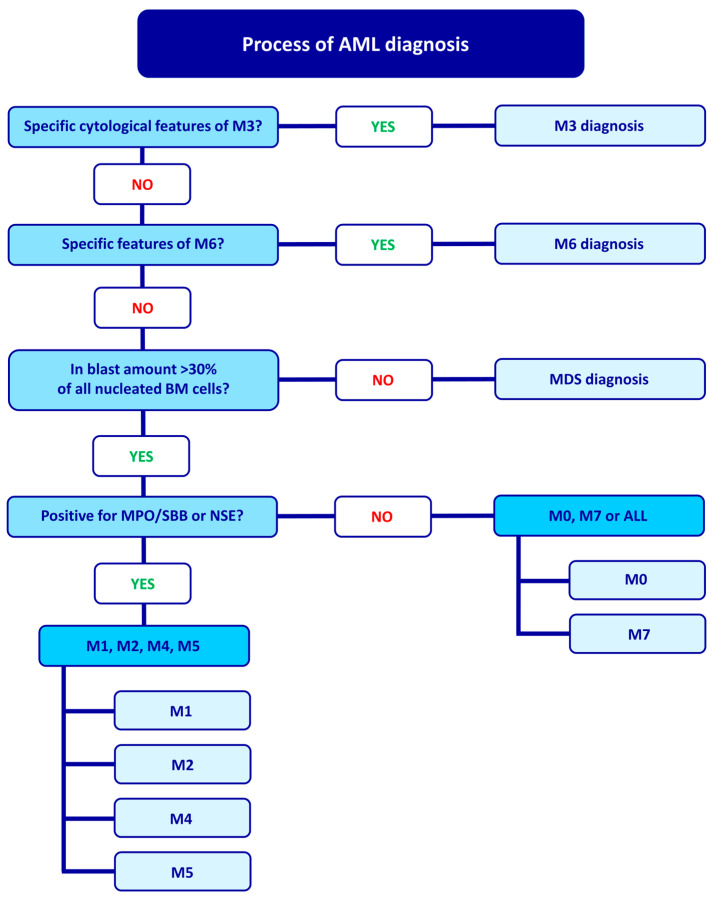
Immortalized cell lines are widely used in vitro tools in oncology and hematology research. While these cell lines represent artificial systems and may accumulate genetic aberrations with each passage, they are still considered valuable models for pilot, preliminary, and screening studies. Despite their limitations, cell lines are cost-effective and provide repeatable and comparable results. Choosing the appropriate cell line for acute myeloid leukemia (AML) research is crucial for obtaining reliable and relevant results. Several factors should be considered when selecting a cell line for AML research, such as specific markers and genetic abnormalities associated with different subtypes of AML. It is also essential to evaluate the karyotype and mutational profile of the cell line, as these can influence the behavior and response to the treatment of the cells. In this review, we evaluate immortalized AML cell lines and discuss the issues surrounding them concerning the revised World Health Organization and the French-American-British classifications.
Keywords: AML cell lines; AML subtypes; CD markers; FAB; WHO; genetic rearrangements.
Conflict of interest statement
The authors declare no conflict of interest.
Figures


References
-
- Bennett J.M., Catovsky D., Daniel M.T., Flandrin G., Galton D.A., Gralnick H.R., Sultan C. Proposed Revised Criteria for the Classification of Acute Myeloid Leukemia. A Report of the French-American-British Cooperative Group. Ann. Intern. Med. 1985;103:620–625. doi: 10.7326/0003-4819-103-4-620. - DOI - PubMed
-
- Naeim F., Rao P.N. Chapter 11—Acute Myeloid Leukemia. In: Naeim F., Rao P.N., Grody W.W., editors. Hematopathology. Academic Press; Oxford, UK: 2008. pp. 207–255. - DOI
-
- Ladines-Castro W., Barragán-Ibañez G., Luna-Pérez M.A., Santoyo-Sánchez A., Collazo-Jaloma J., Mendoza-García E., Ramos-Peñafiel C.O. Morphology of Leukaemias. Rev. Médica Hosp. Gen. México. 2016;79:107–113. doi: 10.1016/j.hgmx.2015.06.007. - DOI
-
- Khoury J.D., Solary E., Abla O., Akkari Y., Alaggio R., Apperley J.F., Bejar R., Berti E., Busque L., Chan J.K.C., et al. The 5th Edition of the World Health Organization Classification of Haematolymphoid Tumours: Myeloid and Histiocytic/Dendritic Neoplasms. Leukemia. 2022;36:1703–1719. doi: 10.1038/s41375-022-01613-1. - DOI - PMC - PubMed
Publication types
MeSH terms
LinkOut - more resources
Full Text Sources
Other Literature Sources
Medical
Research Materials
