. 2023 Mar 14;24(6):5553.
doi: 10.3390/ijms24065553.

Synergistic Interaction of the Class IIa HDAC Inhibitor CHDI0039 with Bortezomib in Head and Neck Cancer Cells

Affiliations

Synergistic Interaction of the Class IIa HDAC Inhibitor CHDI0039 with Bortezomib in Head and Neck Cancer Cells

Christian Schrenk et al. Int J Mol Sci. .

Abstract

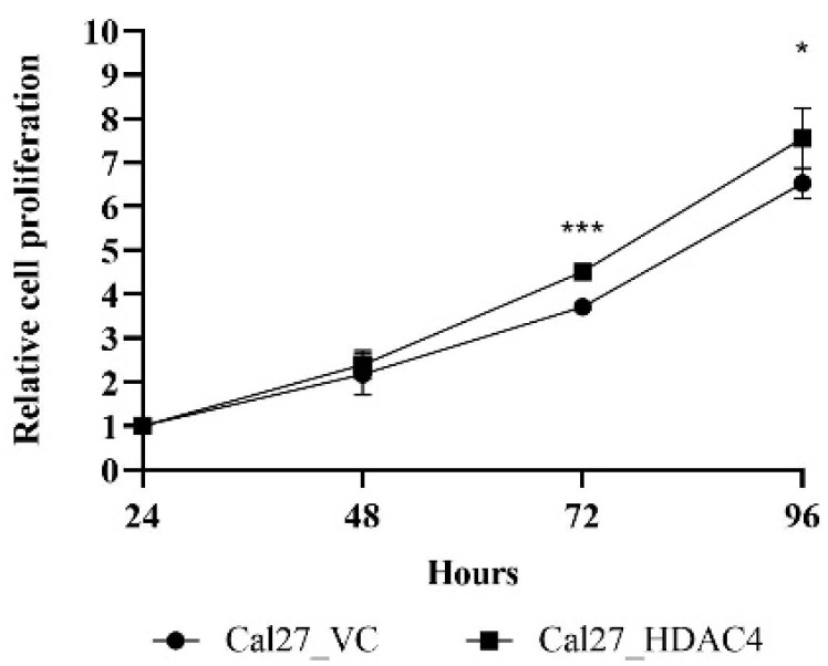
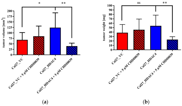
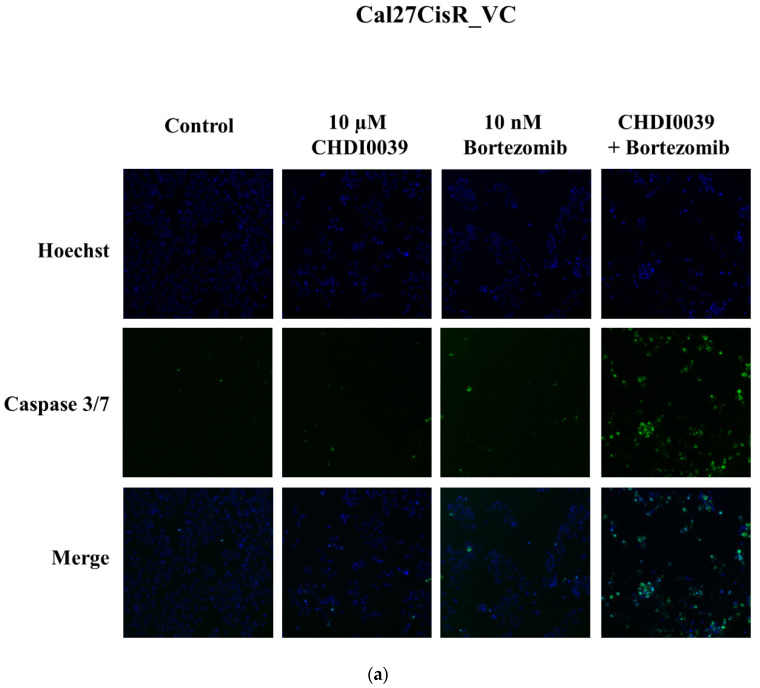
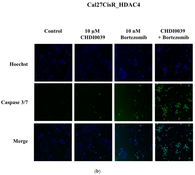
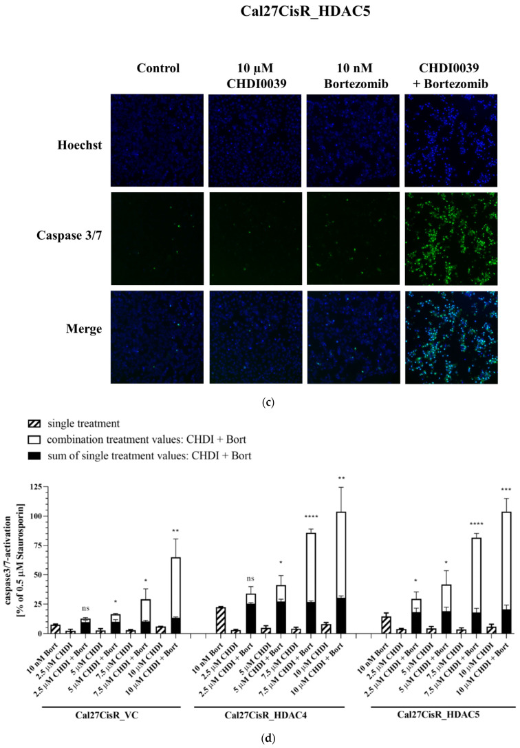
In contrast to class I/IIb/pan histone deacetylase inhibitors (HDACi), the role of class IIa HDACi as anti-cancer chemosensitizing agents is less well understood. Here, we studied the effects of HDAC4 in particular and the class IIa HDACi CHDI0039 on proliferation and chemosensitivity in Cal27 and cisplatin-resistant Cal27CisR head and neck squamous cell cancer (HNSCC). HDAC4 and HDAC5 overexpression clones were generated. HDAC4 overexpression (Cal27_HDAC4) increased proliferation significantly compared to vector control cells (Cal27_VC). Chicken chorioallantoic membrane (CAM) studies confirmed the in vitro results: Cal27_HDAC4 tumors were slightly larger than tumors from Cal27_VC, and treatment with CHDI0039 resulted in a significant decrease in tumor size and weight of Cal27_HDAC4 but not Cal27_VC. Unlike class I/pan-HDACi, treatment with CHDI0039 had only a marginal impact on cisplatin cytotoxicity irrespective of HDAC4 and HDAC5 expression. In contrast, the combination of CHDI0039 with bortezomib was synergistic (Chou-Talalay) in MTT and caspase 3/7 activation experiments. RNAseq indicated that treatment with CHDI0039 alters the expression of genes whose up- or downregulation is associated with increased survival in HNSCC patients according to Kaplan-Meier data. We conclude that the combination of class IIa HDACi with proteasome inhibitors constitutes an effective treatment option for HNSCC, particularly for platinum-resistant cancers.

Keywords: CHDI0039; HDAC inhibitor; HDAC4; HDAC5; bortezomib; class IIa histone deacetylase; head and neck cancer.


Conflict of interest statement

The authors declare no conflict of interest.

Figures

Figure 1
Figure 1
(a) HDAC isoform gene expression in Cal27_VC and Cal27_HDAC4. Data shown are RPKM (reads per kilobase per million mapped reads) expression values normalized from three independent RNA sequencing experiments. The white bars represent the vector control cell line Cal27_VC, the black bars represent the HDAC4 overexpression clone Cal27_HDAC4. (b) Representative Western blot analysis of HDAC protein expression in vector control (VC), HDAC4 and HDAC5 overexpression clones of sensitive Cal27 and cisplatin resistant Cal27CisR cells. (c) Cisplatin concentration effect curves and IC50 values for Cal27_VC and the corresponding cisplatin resistant subline Cal27CisR_VC measured by MTT assay. Resistance factor (RF) is 4-fold.
Figure 4
Figure 4
Median TPM (transcripts per million) values of SERPINB2, TAGLN, and HIST1H2BD in tumor versus normal head–neck tissue. A total of 519 HNSCC tissues and 44 normal tissues from the GEPIA database were used for this analysis. Data were taken from http://gepia.cancer-pku.cn, accessed on 12 December 2022 [33].
Figure 2
Figure 2
Effects of HDAC4 overexpression on cell proliferation analyzed over a time period up to 96 h by MTT assay. The value for 24 h was defined as a starting point with a relative proliferation set as 1. Values represent means ± SD of at least three independent experiments. Significance was calculated by t-test (* p ≤ 0.05, *** p ≤ 0.001).
Figure 3
Figure 3
Effects of HDAC4 overexpression and treatment with 5 µM CHDI0039 on tumor volume (a) and weight (b) in the chorioallantoic membrane (CAM) model. Tumors were seeded on the CAM and grown for 7 days. Treatment with CHDI0039 or buffer control was conducted at day 2 and day 4 after seeding. Values represent means ± SD of at least three independent experiments. Significance was calculated by t-test (ns: not significant; * p ≤ 0.05; ** p ≤ 0.01).
Figure 5
Figure 5
Representative fluorescent images of various Cal27CisR cell clones either untreated (control) or treated with 10 µM CHDI0039, or 10 nM bortezomib, or the combination thereof for 24 h. Hoechst 33342 (“Hoechst”, blue color) was used as nuclear staining and CellEvent Caspase-3/7 green detection reagent (green color) was used for caspase 3/7 activation. (a) Cal27CisR_VC cells. (b) Cal27CisR_HDAC4 cells. (c) Cal27CisR_HDAC5 cells. (d) Caspase 3/7 activation by single treatment and combinations of different concentrations of CHDI0039 (CHDI) and 10 nM bortezomib (Bort) in Cal27CisR_VC, Cal27CisR_HDAC4, and Cal27CisR_HDAC5. Cells were incubated with CHDI0039, bortezomib, or a combination thereof for 24 h. An amount of 0.5 µM Staurosporin was incubated for 8 h and served as positive control. Caspase 3/7 activation was analyzed by ArrayScan XTI. Data are the mean ± SD of two experiments, each with three replicates. Statistical analysis to compare the caspase 3/7 activation of the indicated treatments was performed using t-test. ns p > 0.05, * (p ≤ 0.05), ** (p ≤ 0.01), *** (p ≤ 0.001), **** (p ≤ 0.0001).
Figure 5
Figure 5
Representative fluorescent images of various Cal27CisR cell clones either untreated (control) or treated with 10 µM CHDI0039, or 10 nM bortezomib, or the combination thereof for 24 h. Hoechst 33342 (“Hoechst”, blue color) was used as nuclear staining and CellEvent Caspase-3/7 green detection reagent (green color) was used for caspase 3/7 activation. (a) Cal27CisR_VC cells. (b) Cal27CisR_HDAC4 cells. (c) Cal27CisR_HDAC5 cells. (d) Caspase 3/7 activation by single treatment and combinations of different concentrations of CHDI0039 (CHDI) and 10 nM bortezomib (Bort) in Cal27CisR_VC, Cal27CisR_HDAC4, and Cal27CisR_HDAC5. Cells were incubated with CHDI0039, bortezomib, or a combination thereof for 24 h. An amount of 0.5 µM Staurosporin was incubated for 8 h and served as positive control. Caspase 3/7 activation was analyzed by ArrayScan XTI. Data are the mean ± SD of two experiments, each with three replicates. Statistical analysis to compare the caspase 3/7 activation of the indicated treatments was performed using t-test. ns p > 0.05, * (p ≤ 0.05), ** (p ≤ 0.01), *** (p ≤ 0.001), **** (p ≤ 0.0001).

References

    1. Asfaha Y., Schrenk C., Alves Avelar L.A., Hamacher A., Pflieger M., Kassack M.U., Kurz T. Recent advances in class IIa histone deacetylases research. Bioorg. Med. Chem. 2019;27:115087. doi: 10.1016/j.bmc.2019.115087. - DOI - PubMed
    1. Eckschlager T., Plch J., Stiborova M., Hrabeta J. Histone Deacetylase Inhibitors as Anticancer Drugs. Int. J. Mol. Sci. 2017;18:1414. doi: 10.3390/ijms18071414. - DOI - PMC - PubMed
    1. Harada T., Hideshima T., Anderson K.C. Histone deacetylase inhibitors in multiple myeloma: From bench to bedside. Int. J. Hematol. 2016;104:300–309. doi: 10.1007/s12185-016-2008-0. - DOI - PubMed
    1. Yang F., Zhao N., Ge D., Chen Y. Next-generation of selective histone deacetylase inhibitors. RSC Adv. 2019;9:19571–19583. doi: 10.1039/C9RA02985K. - DOI - PMC - PubMed
    1. Kaletsch A., Pinkerneil M., Hoffmann M.J., Jaguva Vasudevan A.A., Wang C., Hansen F.K., Wiek C., Hanenberg H., Gertzen C., Gohlke H., et al. Effects of novel HDAC inhibitors on urothelial carcinoma cells. Clin. Epigenet. 2018;10:100. doi: 10.1186/s13148-018-0531-y. - DOI - PMC - PubMed

MeSH terms