Review
. 2023 Mar 15;24(6):5618.
doi: 10.3390/ijms24065618.

Dopamine Transmission Imbalance in Neuroinflammation: Perspectives on Long-Term COVID-19

Affiliations
Review

Dopamine Transmission Imbalance in Neuroinflammation: Perspectives on Long-Term COVID-19

Maria Mancini et al. Int J Mol Sci. .

Abstract

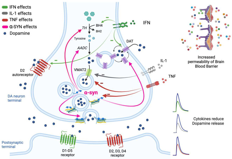
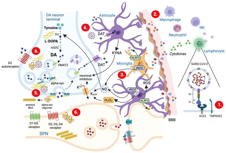
Dopamine (DA) is a key neurotransmitter in the basal ganglia, implicated in the control of movement and motivation. Alteration of DA levels is central in Parkinson's disease (PD), a common neurodegenerative disorder characterized by motor and non-motor manifestations and deposition of alpha-synuclein (α-syn) aggregates. Previous studies have hypothesized a link between PD and viral infections. Indeed, different cases of parkinsonism have been reported following COVID-19. However, whether SARS-CoV-2 may trigger a neurodegenerative process is still a matter of debate. Interestingly, evidence of brain inflammation has been described in postmortem samples of patients infected by SARS-CoV-2, which suggests immune-mediated mechanisms triggering the neurological sequelae. In this review, we discuss the role of proinflammatory molecules such as cytokines, chemokines, and oxygen reactive species in modulating DA homeostasis. Moreover, we review the existing literature on the possible mechanistic interplay between SARS-CoV-2-mediated neuroinflammation and nigrostriatal DAergic impairment, and the cross-talk with aberrant α-syn metabolism.

Keywords: Parkinson’s disease; SARS-CoV-2; alpha-synuclein; cytokines; dopamine; dopamine release; glia; interleukins; long-COVID; neuroinflammation; post-acute sequelae.


Conflict of interest statement

The authors declare no conflict of interest.

Figures

Figure 1
Figure 1
Potential mechanisms of cytokine effects on DA neurotransmission. Simplified representation. Cytokines released from peripheral immune cells or those produced in the brain by activated astrocytes and microglia lead to decreased concentrations of synaptic DA by effect on its synthesis and release. DA is synthesized in the cytoplasm by the action of tyrosine hydroxylase (TH) and amino acid decarboxylase (AADC). Once synthesized, DA is immediately sequestered into vesicles by the vesicular monoamine transporter 2 (VMAT2) and then released into the synaptic cleft where it binds to its postsynaptic receptors. DA signaling at synapse is terminated by degradation or reuptake of DA via the DA transporter (DAT). Inflammatory cytokines (e.g., IFN) may in fact impair the availability of DA by contributing to the oxidation of BH4, the cofactor required for the conversion of tyrosine to L-DOPA. They may also decrease the expression or function of VMAT2 (e.g., IL-1 and TNF) and/or increase the expression or function of DAT (e.g., IFN). The disruption in DA homeostasis induced by cytokines (e.g., TNF) may also be consequence of α-syn changes; in fact, α-syn is involved in the regulation of TH and AADC activity, in the regulation of synaptic vesicle fusion into the synaptic cleft, and in the trafficking of DAT to the cell surface. Finally, an inhibitory effect on release is also caused by a reduction in D2 receptors (e.g., IFN).
Figure 2
Figure 2
Schematic working model illustrating various proposed cellular mechanisms for how altered DA release could take place following SARS-CoV-2 infection. (1.) The spike protein of SARS-Cov-2 virus, by binding the protein angiotensin-converting enzyme 2 (ACE2), and under the participation of type II transmembrane serine protease receptors (TMPRSS2) [41], allows the virus to enter the cell and replicate itself using the RNA and protein synthesis machinery of the host. (2.) SARS-CoV-2 virus, via its spike protein, can activate peripheral immune cells (macrophages, T lymphocytes, NK cells, neutrophils), which release cytokines and chemokines. Cytokines, in turn, increase the permeability of the brain–blood barrier (BBB) and gain access to neurons, astrocytes, and microglial cells. (3.) Activated astrocytes and microglia synthesize other cytokines and produce many proinflammatory molecules, including hydrogen peroxide (H2O2), nitric oxide (NO), and kynurenic acid (KYNA), which cause neuronal inhibition and thereby reduce DA release. Also, microglia cells when activated express NLRP3 (NLR family pyrin domain containing 3) inflammasome, a complex of intracellular proteins which are involved in the maturation of proinflammatory cytokines (see text for details). (4.) The viral particles can enter the CNS (see text for details) and cause a direct activation of astrocytes and microglia that contribute to the inflammatory response in the CSF. (5.) The interaction between viral particles and α-syn causes impairment in vesicle docking and recycling and prevents the loading of newly synthesized and taken up DA into vesicle. (6.) After SARS-CoV-2 invasion, α-syn displays an increased trend to form oligomers and fibrils and to spread up throughout the synapse. Consequently, α-syn has an increased potential to affect DA transmission after viral invasion, compared to what was already described in Figure 1. Thus, the combined action of α-syn dysfunction and the increased inflammatory molecules could drive DA release alterations and participate in the onset of PD. CNS = Central Nervous System. CSF = Cerebral Spinal Fluid DAT = Dopamine Transporter. iNOS = inducible Nitric Oxide Synthase. ROS = Reactive Oxygen Species. SPN = Spiny Projection Neurons.

Similar articles

Cited by

References

    1. Greenhalgh A.D., David S., Bennett F.C. Immune cell regulation of glia during CNS injury and disease. Nat. Rev. Neurosci. 2020;21:139–152. doi: 10.1038/s41583-020-0263-9. - DOI - PubMed
    1. Fani Maleki A., Rivest S. Innate immune cells: Monocytes, monocyte-derived macrophages and microglia as therapeutic target for Alzheimer’s disease and multiple sclerosis. Front. Cell Neurosci. 2019;13:355. doi: 10.3389/fncel.2019.00355. - DOI - PMC - PubMed
    1. Vainchtein I.D., Molofsky A.V. Astrocytes and microglia: In sickness and in health. Trends Neurosci. 2020;43:144–154. doi: 10.1016/j.tins.2020.01.003. - DOI - PMC - PubMed
    1. Xanthos D.N., Sandkühler J. Neurogenic inflammation: Inflammatory CNS reactions in response to neuronal activity. Nat. Rev. Neurosci. 2014;15:43–53. doi: 10.1038/nrn3617. - DOI - PubMed
    1. Habbas S., Santello M., Becker D., Stubbe H., Zappia G., Liaudet N., Klaus F.R., Kollias G., Fontana A., Pryce C.R., et al. Neuroinflammatory TNFα impairs memory via astrocyte signaling. Cell. 2015;163:1730–1741. doi: 10.1016/j.cell.2015.11.023. - DOI - PubMed