. 2023 Mar 16;24(6):5666.
doi: 10.3390/ijms24065666.

Functional and Phenotypic Characterisations of Common Syngeneic Tumour Cell Lines as Estrogen Receptor-Positive Breast Cancer Models

Affiliations

Functional and Phenotypic Characterisations of Common Syngeneic Tumour Cell Lines as Estrogen Receptor-Positive Breast Cancer Models

Maria Lambouras et al. Int J Mol Sci. .

Abstract

Estrogen receptor-positive breast cancers (ER+ BCas) are the most common form of BCa and are increasing in incidence, largely due to changes in reproductive practices in recent decades. Tamoxifen is prescribed as a component of standard-of-care endocrine therapy for the treatment and prevention of ER+ BCa. However, it is poorly tolerated, leading to low uptake of the drug in the preventative setting. Alternative therapies and preventatives for ER+ BCa are needed but development is hampered due to a paucity of syngeneic ER+ preclinical mouse models that allow pre-clinical experimentation in immunocompetent mice. Two ER-positive models, J110 and SSM3, have been reported in addition to other tumour models occasionally shown to express ER (for example 4T1.2, 67NR, EO771, D2.0R and D2A1). Here, we have assessed ER expression and protein levels in seven mouse mammary tumour cell lines and their corresponding tumours, in addition to their cellular composition, tamoxifen sensitivity and molecular phenotype. By immunohistochemical assessment, SSM3 and, to a lesser extent, 67NR cells are ER+. Using flow cytometry and transcript expression we show that SSM3 cells are luminal in nature, whilst D2.0R and J110 cells are stromal/basal. The remainder are also stromal/basal in nature; displaying a stromal or basal Epcam/CD49f FACS phenotype and stromal and basal gene expression signatures are overrepresented in their transcript profile. Consistent with a luminal identity for SSM3 cells, they also show sensitivity to tamoxifen in vitro and in vivo. In conclusion, the data indicate that the SSM3 syngeneic cell line is the only definitively ER+ mouse mammary tumour cell line widely available for pre-clinical research.

Keywords: breast cancer cell line; estrogen receptor-positive; mouse models; syngeneic.


Conflict of interest statement

The authors declare no conflict of interest.

Figures

Figure 1
Figure 1
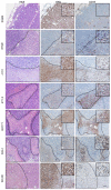
Erα expression in a collection of mouse mammary tumours. Immunohistochemical staining of Erα in epithelial tumour cells of various syngeneic mouse models. The only two cell lines producing tumours with nuclear Erα expression are the 67NR and SSM3 cells. Control: normal mouse mammary gland. Scale bar = 50 µm.
Figure 2
Figure 2
Distribution of Erα positive and CD45-positive cells in syngeneic tumours. Immunohistochemical staining of Erα and CD45 in tumours derived from different syngeneic mouse mammary tumours. ERα staining in the cytoplasm or, in the case of the J110 cell line, in the nucleus, shows concurrent staining with CD45, thus representing immune cell infiltrates. Dotted lines delineate areas of immune infiltration separate from epithelial cell counterparts. Scale bar SSM3 = 100 µm. Scale bar for all other tumours = 50 µm.
Figure 3
Figure 3
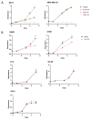
Response of syngeneic cell lines to tamoxifen treatment in vitro. Cells were incubated with either vehicle or 0.1 µM–10 µM 4-hydroxytamoxifen and proliferation was measured over 5–6 days (mean ± SEM of three to six replicate samples). (A) Response of commonly used human xenograft cell lines. MCF7 represents ER+ cells and MDA-MB-231 represents ER-cells. Both respond as expected. (B) Response of mouse mammary tumour lines. Tam, tamoxifen. Data shown as mean ± SEM. *** p ≤ 0.001, ** p ≤ 0.01.
Figure 4
Figure 4
Response of SSM3 cell line to tamoxifen in vivo. (A) SSM3 tumour growth over time in tamoxifen-treated vs vehicle control-treated 129SvEv mice. 129SvEv mice inoculated with 100,000 SSM3 ER+ tumour cells and treated daily with vehicle control (n = 10) or 1 mg tamoxifen citrate (n = 7) once tumours reached 200 mm3. (B) Change in body weight from baseline of mice injected with SSM3 cells and treated with tamoxifen compared to vehicle. Data are presented as mean ± SEM. Data were analysed with a mixed-effects two-way ANOVA and Sidak’s multiple comparisons test. **** p ≤ 0.0001, *** p ≤ 0.001, ** p ≤ 0.01, * p ≤ 0.05.
Figure 5
Figure 5
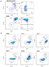
FACS profiles of syngeneic cell lines. Cells were grown in a culture and then assessed using flow cytometry markers for mammary epithelial cell subpopulations. FACS plots shown have already been gated to remove doublets, dead cells (PI positive) and lineage cells (CD45-, CD31-, TER119-). (A) Plots show a normal mouse mammary epithelial cell profile. EpCAM and CD49f delineate luminal, basal and stromal cells. Specific luminal cell populations can be further resolved using CD49b and Sca1. SSM3 cells are shown here as a luminal ER+ cell line. (B) Resolution of the epithelial cell subpopulations in six other mammary tumour lines. (LM, luminal mature; LP, luminal progenitor).
Figure 6
Figure 6
Molecular assessment of syngeneic cell lines and expression of cell-specific gene signatures. (A) Heatmap of genes in the MaSC, luminal progenitor and luminal mature gene signatures [16] and a stromal gene signature [15] (BD) Gene signature scores calculated using MaSC, luminal progenitor and luminal mature genes [16] and stromal gene signature [15] in (B) J110 and SSM3 cells, (C) 4T1.2, 67NR, EO771 cells (GSE42272) and (D) D2A1 and D2.0R cells (GSE172882). Differences in the mean signature scores were calculated using student’s t-test. **** ≤ p 0.0001, *** ≤ p 0.001, ** p ≤ 0.01, * p ≤ 0.05.

References

    1. Cuzick J., Sestak I., Cawthorn S., Hamed H., Holli K., Howell A., Forbes J.F., Investigators I.-I. Tamoxifen for prevention of breast cancer: Extended long-term follow-up of the IBIS-I breast cancer prevention trial. Lancet Oncol. 2015;16:67–75. doi: 10.1016/S1470-2045(14)71171-4. - DOI - PMC - PubMed
    1. Normanno N., Di Maio M., De Maio E., De Luca A., De Matteis A., Giordano A., Perrone F. Mechanisms of endocrine resistance and novel therapeutic strategies in breast cancer. Endocr.-Relat. Cancer. 2005;12:721–747. doi: 10.1677/erc.1.00857. - DOI - PubMed
    1. Smith S.G., Sestak I., Forster A., Partridge A., Side L., Wolf M., Horne R., Wardle J., Cuzick J. Factors affecting uptake and adherence to breast cancer chemoprevention: A systematic review and meta-analysis. Ann. Oncol. 2016;27:575–590. doi: 10.1093/annonc/mdv590. - DOI - PMC - PubMed
    1. Donnelly L., Evans D., Wiseman J., Fox J., Greenhalgh R., Affen J., Juraskova I., Stavrinos P., Dawe S., Cuzick J. Uptake of tamoxifen in consecutive premenopausal women under surveillance in a high-risk breast cancer clinic. Br. J. Cancer. 2014;110:1681–1687. doi: 10.1038/bjc.2014.109. - DOI - PMC - PubMed
    1. DeCensi A., Thorat M.A., Bonanni B., Smith S.G., Cuzick J. Barriers to preventive therapy for breast and other major cancers and strategies to improve uptake. Ecancermedicalscience. 2015;9:595. doi: 10.3332/ecancer.2015.595. - DOI - PMC - PubMed