. 2023 Mar 16;24(6):5692.
doi: 10.3390/ijms24065692.

Adipose-Derived Stem Cells in Reinforced Collagen Gel: A Comparison between Two Approaches to Differentiation towards Smooth Muscle Cells

Affiliations

Adipose-Derived Stem Cells in Reinforced Collagen Gel: A Comparison between Two Approaches to Differentiation towards Smooth Muscle Cells

Elena Filova et al. Int J Mol Sci. .

Abstract

Scaffolds made of degradable polymers, such as collagen, polyesters or polysaccharides, are promising matrices for fabrication of bioartificial vascular grafts or patches. In this study, collagen isolated from porcine skin was processed into a gel, reinforced with collagen particles and with incorporated adipose tissue-derived stem cells (ASCs). The cell-material constructs were then incubated in a DMEM medium with 2% of FS (DMEM_part), with added polyvinylalcohol nanofibers (PVA_part sample), and for ASCs differentiation towards smooth muscle cells (SMCs), the medium was supplemented either with human platelet lysate released from PVA nanofibers (PVA_PL_part) or with TGF-β1 + BMP-4 (TGF + BMP_part). The constructs were further endothelialised with human umbilical vein endothelial cells (ECs). The immunofluorescence staining of alpha-actin and calponin, and von Willebrand factor, was performed. The proteins involved in cell differentiation, the extracellular matrix (ECM) proteins, and ECM remodelling proteins were evaluated by mass spectrometry on day 12 of culture. Mechanical properties of the gels with ASCs were measured via an unconfined compression test on day 5. Gels evinced limited planar shrinkage, but it was higher in endothelialised TGF + BMP_part gel. Both PVA_PL_part samples and TGF + BMP_part samples supported ASC growth and differentiation towards SMCs, but only PVA_PL_part supported homogeneous endothelialisation. Young modulus of elasticity increased in all samples compared to day 0, and PVA_PL_part gel evinced a slightly higher ratio of elastic energy. The results suggest that PVA_PL_part collagen construct has the highest potential to remodel into a functional vascular wall.

Keywords: adipose tissue-derived stem cells; collagen particles; endothelial cells; extracellular matrix; gel reinforcement; remodelling; stem cells differentiation; tissue engineering; vascular patches.


Conflict of interest statement

The authors declare no conflict of interest.

Figures
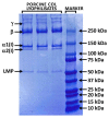
Figure 1
Figure 1
Polypeptide pattern of COL I type isolated from porcine skin.
Figure 2
Figure 2
FTIR spectrum of COL I type isolated from pork skin.
Figure 3
Figure 3
SEM image of electrospun collagen nano/submicrofibers (A; mag. 50,000×) and collagen particles prepared via homogenisation of collagen electrospun layers (B; mag. 500×).
Figure 4
Figure 4
(A) SEM image of electrospun PVA with incorporated platelet lysate (PVA_PL), scale bar 10 µm; (B) histogram of the fiber diameter distribution in PVA_PL material; (C) 10% SDS-PAGE documenting the protein release from PVA_PL after 1, 2, 4, 6, 8, 24, 72 and 168 h in PBS; PVA material without incorporated proteins served as negative control (NC), Coomassie blue staining.
Figure 5
Figure 5
Crystallinity analysis (DSC) of PVA (A) and PVA_PL (B).
Figure 6
Figure 6
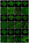
Living ASCs, stained with CellTrackerTM Green CMFDA dye, in collagen gel with collagen particles on day 1 (AD,A`D`), on day 7 (EH,E`H`) and on day 14 (IL,I`L`). The cells were cultured in DMEM with 2% of FS (A,A`,E,E`,I,I`), in medium with added PVA nanomats (B,B`,F,F`,J,J`) or with added PVA_PL nanomats (C,C`,G,G`,K,K`) or in medium with TGF-β1 + BPM-4 (D,D`,H,H`,L,L`). Dragonfly 503 (Andor)—spinning disk confocal microscope, obj. × 10, 3D vizualisation (AL) and maximum projection (A`L`), scale bar = 200 µm, image size 820 × 820 × 500 µm.
Figure 7
Figure 7
(A) Densities of ASCs cultured in collagen gels with particles on days 1, 7, and 14, and density of both ASCs and ECs in endothelialised gels on day 14; (B) gel area. Data are expressed as mean ± S.D. Statistical significance is considered * for p-value < 0.05.
Figure 8
Figure 8
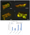
Immunofluorescence staining of alpha-actin (red) and calponin (green) in ASCs in collagen gels with collagen particles on day 7. The cells were cultured in DMEM with 2% of FS (A), in medium with added PVA nanomats (B), with added PVA_PL nanomats (C) or in medium supplemented with TGF-β1 + BPM-4 (D), scale bar = 800 µm. Percentage of calponin+ cells in collagen gels on days 7 and 14 (E). Cell nuclei were counterstained with DAPI. Zeiss Z.1 lightsheet microscope, obj. × 10 for excitation, obj. × 20 for detection, zoom × 0.4 tile scans. Data are expressed as mean ± S.D. Statistical significance is considered for * p-value < 0.05 in comparison with the sample of the same number, and is depicted above the graph columns.
Figure 9
Figure 9
Immunofluorescence staining of fibronectin (green, AD,A*D*), type I collagen (red, A`D`,A*D*), second harmonic generation signal of fibrous collagen (purple, A``D``,A*D*) in ASCs in collagen gel. The cells were cultured in DMEM with 2% of FS (A,A`,A``,A*), in medium with added PVA nanomats (B,B`,B``,B*), with added PVA_PL nanomats (C,C`,C``,C*) or in medium with TGF-β1 + BPM-4 (D,D`,D``,D*) for 7 days. Zeiss, LSM 780 microscope system, obj. × 63×, image size 135 µm × 67 µm, scale bar = 25 µm.
Figure 10
Figure 10
Immunofluorescence staining of von Willebrand factor in ECs (green) and alpha-actin (red) in ASCs in collagen gel reinforced with collagen particles cultured in DMEM with 2% of FS (A), with added PVA nanomats (B), with PVA_PL nanomats (C) or in medium with TGF-β1 + BMP-4 (D) for 8 days and, subsequently, in EGM-2 medium, for 6 days. The cell nuclei are counterstained with Hoechst, Bruker Ultima, obj. × 25, scale bar = 50 µm.
Figure 11
Figure 11
Initial Young modulus of elasticity of reinforced collagen gel with ASCs immediately after preparation, i.e., DMEM_D0, and on day 5 after seeding, i.e., DMEM_part, PVA_part, PVA_PL_part and TGF + BMP_part. box plot diagram, * denotes statistically significant differences (Student’s t-test, 0.05).
Figure 12
Figure 12
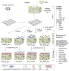
Scheme of the experiments.

References

    1. World Health Organisation Cardiovascular Diseases (CVDs) [(accessed on 8 March 2021)]. Available online: https://www.who.int/news-room/fact-sheets/detail/cardiovascular-diseases...
    1. Moore M.J., Tan R.P., Yang N., Rnjak-Kovacina J., Wise S.G. Bioengineering artificial blood vessels from natural materials. Trends Biotechnol. 2022;40:693–707. doi: 10.1016/j.tibtech.2021.11.003. - DOI - PubMed
    1. Ebert N., McGinnis M., Johnson W., Kuhn E.M., Mitchell M.E., Tweddell J.S., Woods R.K. Comparison of Patch Materials for Pulmonary Artery Reconstruction. Semin. Thorac. Cardiovasc. Surg. 2021;33:459–465. doi: 10.1053/j.semtcvs.2020.09.011. - DOI - PMC - PubMed
    1. Zhang F., Xie Y., Celik H., Akkus O., Bernacki S.H., King M.W. Engineering small-caliber vascular grafts from collagen filaments and nanofibers with comparable mechanical properties to native vessels. Biofabrication. 2019;11:035020. doi: 10.1088/1758-5090/ab15ce. - DOI - PMC - PubMed
    1. Yao Y., Wang J., Cui Y., Xu R., Wang Z., Zhang J., Wang K., Li Y., Zhao Q., Kong D. Effect of sustained heparin release from PCL/chitosan hybrid small-diameter vascular grafts on anti-thrombogenic property and endothelialisation. Acta Biomater. 2014;10:2739–2749. doi: 10.1016/j.actbio.2014.02.042. - DOI - PubMed

LinkOut - more resources