. 2023 Mar 18;24(6):5800.
doi: 10.3390/ijms24065800.

Characterization of the Involvement of Tumour Necrosis Factor (TNF)-α-Stimulated Gene 6 (TSG-6) in Ischemic Brain Injury Caused by Middle Cerebral Artery Occlusion in Mouse

Affiliations

Characterization of the Involvement of Tumour Necrosis Factor (TNF)-α-Stimulated Gene 6 (TSG-6) in Ischemic Brain Injury Caused by Middle Cerebral Artery Occlusion in Mouse

Chiara Di Santo et al. Int J Mol Sci. .

Abstract

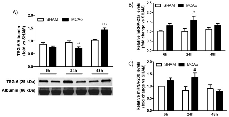
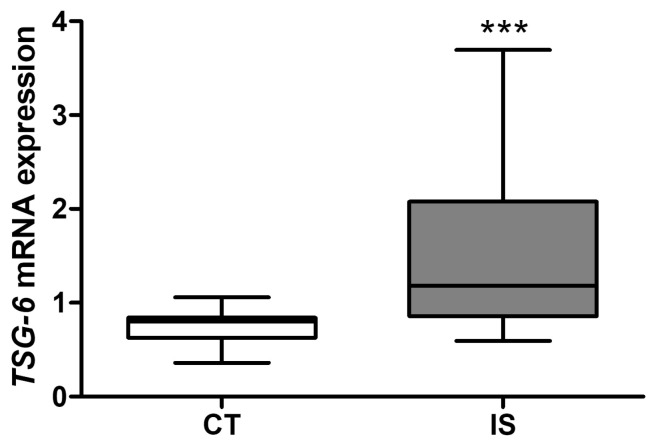
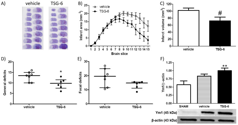
The identification of novel targets to modulate the immune response triggered by cerebral ischemia is crucial to promote the development of effective stroke therapeutics. Since tumour necrosis factor (TNF)-α-stimulated gene 6 (TSG-6), a hyaluronate (HA)-binding protein, is involved in the regulation of immune and stromal cell functions in acute neurodegeneration, we aimed to characterize its involvement in ischemic stroke. Transient middle cerebral artery occlusion (1 h MCAo, followed by 6 to 48 of reperfusion) in mice resulted in a significant elevation in cerebral TSG-6 protein levels, mainly localized in neurons and myeloid cells of the lesioned hemisphere. These myeloid cells were clearly infiltrating from the blood, strongly suggesting that brain ischemia also affects TSG-6 in the periphery. Accordingly, TSG-6 mRNA expression was elevated in peripheral blood mononuclear cells (PBMCs) from patients 48 h after ischemic stroke onset, and TSG-6 protein expression was higher in the plasma of mice subjected to 1 h MCAo followed by 48 h of reperfusion. Surprisingly, plasma TSG-6 levels were reduced in the acute phase (i.e., within 24 h of reperfusion) when compared to sham-operated mice, supporting the hypothesis of a detrimental role of TSG-6 in the early reperfusion stage. Accordingly, systemic acute administration of recombinant mouse TSG-6 increased brain levels of the M2 marker Ym1, providing a significant reduction in the brain infarct volume and general neurological deficits in mice subjected to transient MCAo. These findings suggest a pivotal role of TSG-6 in ischemic stroke pathobiology and underscore the clinical relevance of further investigating the mechanisms underlying its immunoregulatory role.

Keywords: TSG-6; cerebral ischemia; immune system; neurodegeneration; neuroinflammation; stroke.


Conflict of interest statement

The authors declare no conflict of interest. The funders had no role in the design of the study; in the collection, analyses, or interpretation of data; in the writing of the manuscript; or in the decision to publish the results.

Figures

Figure 1
Figure 1
Tumour necrosis factor (TNF)-α-stimulated gene 6 (TSG-6) protein expression is elevated in the brain of mice subjected to transient MCAo. (A) TSG-6 protein expression levels detected by Western blotting in the ipsilateral (ischemic and IPSI) and contralateral (CONTRA) cortex of mice subjected to 1 h MCAo followed by 6, 24 or 48 h of reperfusion or mice subjected to sham surgery (SHAM). Data are expressed as means ± S.E.M.; * p < 0.05 vs. CONTRA 6 h, *** p < 0.001 vs. SHAM 24 h and vs. CONTRA 24 h, § p < 0.05 vs. IPSI 6 h, ** p < 0.01 vs. SHAM 48 h and vs. IPSI 48 h (two-way ANOVA followed by Bonferroni post-test; n = 6–8 mice per experimental group). (B) Representative immunofluorescence images showing colocalization (yellow overlapping in merge panels) of TSG6 (green fluorescence) with NeuN immunopositive neurons (red fluorescence) in the ipsilateral and contralateral cortex of mice subjected to 1 h middle cerebral artery occlusion (MCAo) followed by 24 or 48 h of reperfusion. Nuclei were counterstained with DAPI (blue signal). Scale bar = 150 μm. (C) Representative immunofluorescence images showing colocalization (yellow overlapping in merge panels) of TSG6 (green fluorescence) with Ly6B.2 immunopositive myeloid cells (i.e., granulocytes and monocytes/macrophages, red fluorescence) in the ipsilateral cortex of mice subjected to 1 h MCAo followed by 24 or 48 h of reperfusion. The endothelium was labelled with anti-PECAM1 and the nuclei were counterstained with DAPI (blue signal). Arrows and arrowheads indicate TSG-6 immunopositive myeloid cells in perivascular spaces or infiltrating the brain parenchyma, respectively. Scale bar = 75 μm. (D) Schematic representation of the regional and cellular distribution of TSG-6 immunoreactivity in the brain of mice subjected to 1 h MCAo followed by 24 or 48 h of reperfusion. Drawings were obtained from the typical distribution observed by microscopic examination of brain sections (0.70 mm anterior to Bregma) processed for immunofluorescence, as described in the methods section. NeuN and Ly6B.2 immunopositive cells that express TSG-6 are represented by orange or purple symbolic drawings, respectively.
Figure 2
Figure 2
Transient MCAo modulates plasma levels of TSG-6 protein and of miR-23a and miR-23b. (A) TSG-6 protein, (B) miR-23a or (C) miR-23b relative expression levels in plasma of mice following sham surgery (SHAM) or 1 h MCAo (MCAo) followed by 6, 24 or 48 h of reperfusion. Data are expressed as means ± S.E.M.; ** p < 0.01 vs. SHAM 24 h, *** p < 0.001 vs. SHAM 48 h, # p < 0.05 vs. SHAM 24 h (two-way ANOVA followed by Bonferroni post-test, n = 5–8 mice per experimental group).
Figure 3
Figure 3
TSG-6 mRNA expression is elevated 48 h after ischemic stroke in PBMC from patients. TSG-6 mRNA levels in peripheral blood mononuclear cells (PBMCs) isolated from blood of healthy subjects (CT; n = 13) or patients subjected to ischemic stroke 48 h before (IS; n= 25). Data are shown as median, interquartile range and minimum and maximum values; *** p < 0.01 vs. CT (Mann–Whitney test).
Figure 4
Figure 4
Intravenous acute administration of recombinant TSG-6 significantly reduces brain damage and neurological deficits while promoting cerebral expression of M2-like phenotypes following focal cerebral ischemia in mice. (A) Representative cresyl violet-stained coronal brain sections, (B) brain infarct area and (C) volume in mice subjected to 1 h MCAo followed by 48 h of reperfusion and intravenously injected with 30 μg recombinant mouse TSG-6 or vehicle (100 μL PBS) upon reperfusion. Data are expressed as means ± S.E.M.; # p < 0.05 vs. vehicle (unpaired Student’s t test). Effects of the latter treatment on (D) general and (E) focal neurological deficits caused in mice by 1 h MCAo followed by 48 h of reperfusion. Data are expressed as medians and interquartile ranges; * p < 0.05 vs. PBS (two-tailed Mann–Whitney test); n = 6–7 mice per experimental group. (F) Western blotting analysis of Ym1 protein levels in whole brain homogenates from mice subjected to SHAM surgery or 1 h MCAo followed by 48 h of reperfusion and intravenously injected with TSG-6 or vehicle upon reperfusion. Data are expressed as means ± S.E.M.; ** p < 0.01 vs. SHAM (one-way ANOVA followed by Tukey post-test, n = 5–7 mice per experimental group).

References

    1. Feigin V.L., Stark B.A., Johnson C.O., Roth G.A., Bisignano C., Abady G.G., Abbasifard M., Abbasi-Kangevari M., Abd-Allah F., Abedi V., et al. Global, regional, and national burden of stroke and its risk factors, 1990–2019: A systematic analysis for the Global Burden of Disease Study 2019. Lancet Neurol. 2021;20:795–820. doi: 10.1016/S1474-4422(21)00252-0. - DOI - PMC - PubMed
    1. Thomalla G., Boutitie F., Ma H., Koga M., Ringleb P., Schwamm L.H., Wu O., Bendszus M., Bladin C.F., Campbell B.C.V., et al. Intravenous alteplase for stroke with unknown time of onset guided by advanced imaging: Systematic review and meta-analysis of individual patient data. Lancet. 2020;396:1574–1584. doi: 10.1016/S0140-6736(20)32163-2. - DOI - PMC - PubMed
    1. Ma H., Campbell B.C.V., Parsons M.W., Churilov L., Levi C.R., Hsu C., Kleinig T.J., Wijeratne T., Curtze S., Dewey H.M., et al. Thrombolysis Guided by Perfusion Imaging up to 9 Hours after Onset of Stroke. N. Engl. J. Med. 2019;380:1795–1803. doi: 10.1056/NEJMoa1813046. - DOI - PubMed
    1. Hacke W., Kaste M., Bluhmki E., Brozman M., Dávalos A., Guidetti D., Larrue V., Lees K.R., Medeghri Z., Machnig T., et al. Thrombolysis with Alteplase 3 to 4.5 Hours after Acute Ischemic Stroke. N. Engl. J. Med. 2008;359:1317–1329. doi: 10.1056/NEJMoa0804656. - DOI - PubMed
    1. Shafie M., Yu W. Recanalization Therapy for Acute Ischemic Stroke with Large Vessel Occlusion: Where We Are and What Comes Next? Transl. Stroke Res. 2021;12:369–381. doi: 10.1007/s12975-020-00879-w. - DOI - PMC - PubMed

Substances