. 2023 Mar 19;24(6):5838.
doi: 10.3390/ijms24065838.

Aconitum carmichaelii Debx. Attenuates Heart Failure through Inhibiting Inflammation and Abnormal Vascular Remodeling

Affiliations

Aconitum carmichaelii Debx. Attenuates Heart Failure through Inhibiting Inflammation and Abnormal Vascular Remodeling

Ziwei Xing et al. Int J Mol Sci. .

Abstract

Heart failure (HF) is the most common complication following myocardial infarction, closely associated with ventricular remodeling. Aconitum carmichaelii Debx., a traditional Chinese herb, possesses therapeutic effects on HF and related cardiac diseases. However, its effects and mechanisms on HF-associated cardiac diseases are still unclear. In the present study, a water extraction of toasted Aconitum carmichaelii Debx. (WETA) was verified using UPLC-Q/TOF-MS. The heart function of HF rats was assessed by echocardiography and strain analysis, and myocardial injury was measured by serum levels of CK-MB, cTnT, and cTnI. The pathological changes of cardiac tissues were evaluated by 2,3,5-triphenyltetrazolium chloride (TTC) staining, hematoxylin and eosin (H&E) staining, and Masson's trichrome staining. Additionally, the levels of inflammation-related genes and proteins and components related to vascular remodeling were detected by RT-qPCR, Western blot, and immunofluorescence. WETA significantly inhibited the changes in echocardiographic parameters and the increase in heart weight, cardiac infarction size, the myonecrosis, edema, and infiltration of inflammatory cells, collagen deposition in heart tissues, and also mitigated the elevated serum levels of CK-MB, cTnT, and cTnI in ISO-induced rats. Additionally, WETA suppressed the expressions of inflammatory genes, including IL-1β, IL-6, and TNF-α and vascular injury-related genes, such as VCAM1, ICAM1, ANP, BNP, and MHC in heart tissues of ISO-induced HF rats, which were further confirmed by Western blotting and immunofluorescence. In summary, the myocardial protective effect of WETA was conferred through inhibiting inflammatory responses and abnormal vascular remodeling in ISO-treated rats.

Keywords: Aconitum carmichaelii Debx.; Angprotein-2; FOXO1; NF-κB signaling pathway; heart failure; inflammation; vascular remodeling.


Conflict of interest statement

The authors declare that the research was conducted in the absence of any commercial or financial relationships that could be construed as a potential conflict of interest.

Figures
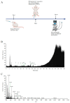
Figure 1
Figure 1
Identification of WETA compounds using LC-MS. (A) The experimental design flow, (B) total ion chromatogram of WETA, (C) MS2 spectrum of WETA. 1, Salsolinol; 2, Karakoline; 3, Isotalatizidine; 4, Napelline; 5, Neoline/Bullatine B; 6, Neoline; 7, Lycoctonine; 8, Fuziline (15-α-Hydroxyneoline); 9, Talatisamine; 10, Benzoylmesaconine; 11, Benzoylaconine; 12, Hypaconitine; 13, Benzoylhypaconine; 14, Aconitine.
Figure 2
Figure 2
WETA improved LV echocardiography parameters in ISO-induced rats. (A) Representative short axis image of the LV, the levels of (B) FS, (C) EF, (D) LVIDs, (E) LVIDd, (F) LVESV, and (G) LVEDV. Values are mean ± SD (n = 6), ** p < 0.01 vs. CON group; ## p < 0.01 vs. ISO group, using one-way ANOVA.
Figure 3
Figure 3
WETA relieved LV segmental myocardial strain in ISO-induced rats. (A) Schematic diagram of the division of the LV into six parts. Representative trace tendency of the LV wall during systole (B) and diastole (C); (D) representative images of RLS, (E) RLS of six segments. (F) Representative images of RRS, (G) RRS of six segments, (H) global longitudinal strain, (I) stroke volume, (J) cardiac output, (K) maximum opposing wall delay. AB, anterior base; AM, anterior middle; AP, anterior apex; PB, posterior base; PM, posterior middle; PA, posterior apex, RRS, regional radial strain, RLS, regional longitudinal strain. Values are mean ± SD (n = 6), * p < 0.05, ** p < 0.01 vs. CON group; # p < 0.05, ## p < 0.01 vs. ISO group, using one-way ANOVA.
Figure 4
Figure 4
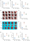
WETA reduced heart deformation and cardiac damage markers. (A) Fasting body weight, (B) heart tissue weight, (C) cardiac tissue index (cardiac weight index (CWI) was calculated according to the following formula: CWI(g/g) = heart tissue weight (g)/body weight (g) × 100), (D) representative heart shape (scale bar = 500 μm), (E) heart area, (F) representative TTC-stained sections (Scale bar = 1 cm), (G) heart infarction area, the serum levels of CK-MB (H), cTNT (I), and cTNI (J). Values are mean ± SD (n = 6), ** p < 0.01 vs. CON group; ## p < 0.01, # p < 0.05 vs. ISO group, using one-way ANOVA.
Figure 5
Figure 5
WETA attenuates histopathological changes. (A) H&E staining images of LV (From left to right, scale bar = 200 μm, 100 μm, 50 μm), (B) Positive cells in the H&E stained slices were normalized and counted as cells/mm2, (C) Masson’s staining images of LV (From left to right, scale bar = 200 μm, 100 μm, 50 μm), and (D) CVF of LV. Scale bar = 200 μm, 200 μm, 50 μm. Black arrows: irregular cross striations of the myocardial tissue. Values are mean ± SD (n = 6), ** p < 0.01 vs. CON group; ## p < 0.01 vs. ISO group, from One-way ANOVA.
Figure 6
Figure 6
WETA attenuated the inflammatory response in heart tissues of ISO-induced rats. (A) Immunofluorescence staining images of CD68 (red), CD31 (green), and nuclei (blue) (scale bar = 40 μm). Mean fluorescence intensity of CD68 (B) and CD31 (C); mRNA levels of IL-1β (D), IL-6 (E), and TNF-α (F). (G) Western blot results of the levels of p-Ikkα/β (H), p-p65 (I), and p-IκBα (J). Values are mean ± SD (n = 6), ** p < 0.01 vs. CON group; ## p < 0.01 vs. ISO group, using one-way ANOVA.
Figure 7
Figure 7
WETA attenuates cardiac myocardial damage. (A) Immunofluorescence staining images of E-selection (red), CD31 (green), and nuclei (blue) (scale bar = 40 μm), (B) mean fluorescence intensity of E-selection, (C) expression of ANP, BNP, MHC, ICAM1, and VCAM1. (D) Western blot results of levels of p-Foxo1 (E), Angpt2 (F), ICAM1 (G) and VCAM1 (H). Values are mean ± SD (n = 6), ** p < 0.01 vs. CON group; ## p < 0.01 vs. ISO group, using one-way ANOVA.

References

    1. Baman J.R., Ahmad F.S. Heart Failure. JAMA. 2020;324:1015. doi: 10.1001/jama.2020.13310. - DOI - PubMed
    1. Heidenreich P.A., Bozkurt B., Aguilar D., Allen L.A., Byun J.J., Colvin M.M., Deswal A., Drazner M.H., Dunlay S.M., Evers L.R., et al. 2022 AHA/ACC/HFSA Guideline for the Management of Heart Failure: Executive Summary: A Report of the American College of Cardiology/American Heart Association Joint Committee on Clinical Practice Guidelines. Circulation. 2022;145:e876–e894. doi: 10.1161/CIR.0000000000001062. - DOI - PubMed
    1. Ezekowitz J.A., O’Meara E., McDonald M.A., Abrams H., Chan M., Ducharme A., Giannetti N., Grzeslo A., Hamilton P.G., Heckman G.A., et al. 2017 Comprehensive Update of the Canadian Cardiovascular Society Guidelines for the Management of Heart Failure. Can. J. Cardiol. 2017;33:1342–1433. doi: 10.1016/j.cjca.2017.08.022. - DOI - PubMed
    1. Anderson T., Hummel S.L., Konerman M.C. Epidemiology, Diagnosis, Pathophysiology, and Initial Approach to Heart Failure with Preserved Ejection Fraction. Cardiol. Clin. 2022;40:397–413. doi: 10.1016/j.ccl.2022.07.001. - DOI - PubMed
    1. Gonzalez A., Schelbert E.B., Diez J., Butler J. Myocardial Interstitial Fibrosis in Heart Failure: Biological and Translational Perspectives. J. Am. Coll. Cardiol. 2018;71:1696–1706. doi: 10.1016/j.jacc.2018.02.021. - DOI - PubMed