. 2023 Mar 19;24(6):5853.
doi: 10.3390/ijms24065853.

Kinetic Characterization and Catalytic Mechanism of N-Acetylornithine Aminotransferase Encoded by slr1022 Gene from Synechocystis sp. PCC6803

Affiliations

Kinetic Characterization and Catalytic Mechanism of N-Acetylornithine Aminotransferase Encoded by slr1022 Gene from Synechocystis sp. PCC6803

Zhi-Min Li et al. Int J Mol Sci. .

Abstract

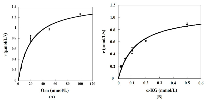
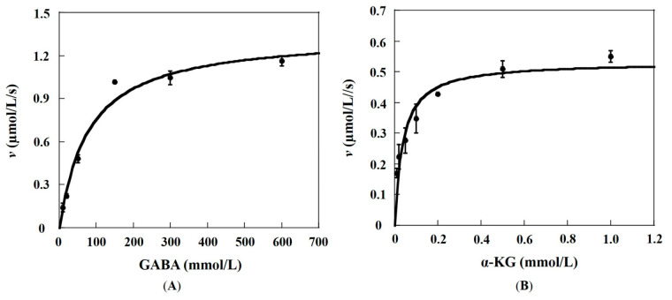
The enzyme encoded by slr1022 gene from Synechocystis sp. PCC6803 was reported to function as N-acetylornithine aminotransferase, γ-aminobutyric acid aminotransferase, and ornithine aminotransferase, which played important roles in multiple metabolic pathways. Among these functions, N-acetylornithine aminotransferase catalyzes the reversible conversion of N-acetylornithine to N-acetylglutamate-5-semialdehyde with PLP as cofactor, which is a key step in the arginine biosynthesis pathway. However, the investigation of the detailed kinetic characteristics and catalytic mechanism of Slr1022 has not been carried out yet. In this study, the exploration of kinetics of recombinant Slr1022 illustrated that Slr1022 mainly functioned as N-acetylornithine aminotransferase with low substrate specificity to γ-aminobutyric acid and ornithine. Kinetic assay of Slr1022 variants and the model structure of Slr1022 with N-acetylornithine-PLP complex revealed that Lys280 and Asp251 residues were the key amino acids of Slr1022. The respective mutation of the above two residues to Ala resulted in the activity depletion of Slr1022. Meanwhile, Glu223 residue was involved in substrate binding and it served as a switch between the two half reactions. Other residues such as Thr308, Gln254, Tyr39, Arg163, and Arg402 implicated a substrate recognition and catalytic process of the reaction. The results of this study further enriched the understanding of the catalytic kinetics and mechanism of N-acetylornithine aminotransferase, especially from cyanobacteria.

Keywords: N-acetylornithine aminotransferase; Synechocystis sp. PCC6803; catalytic mechanism; kinetic characterization; site-directed mutagenesis.


Conflict of interest statement

The authors declare no conflict of interests.

Figures

Figure 1
Figure 1
Kinetic profiles of recombinant Slr1022 protein as OAT. (A): Plot of the initial velocities as function of Orn concentrations, the concentration of α-KG was fixed at 0.5 mmol/L. (B): Plot of the initial velocities as function of α-KG concentrations, the concentration of Orn was fixed at 100 mmol/L. Data are expressed as the mean ± standard deviation, n = 3.
Figure 2
Figure 2
Kinetic profiles of recombinant Slr1022 protein as GABA-AT. (A): Plot of the initial velocities as function of GABA concentrations, the concentration of α-KG was fixed at 0.5 mmol/L. (B): Plot of the initial velocities as function of α-KG concentrations, the concentration of GABA was fixed at 600 mmol/L. Data are expressed as the mean ± standard deviation, n = 3.
Figure 3
Figure 3
Effects of metal ions and temperature on AcOrn transaminase activity of Slr1022. (A): Effects of metal ions on the activity of Slr1022; (B): Temperature effect on the activity of Slr1022. Data are expressed as the mean ± standard deviation, n = 3.
Figure 4
Figure 4
The overlapped cartoon and active site view of Slr1022 model structure. (A): Overlapped cartoon structure of Slr1022. The model structures from RoseTTAFold and SWISS-MODEL were colored as magenta and cyan, respectively. (B): Active site view of Slr1022 model structure generated from RoseTTAFold. The carbon of PLP, AcOrn, and amino acid residues were colored as cyan, magenta, and grey, respectively. Nitrogen, oxygen, and phosphor atoms were colored as blue, red, and orange, respectively. The hydrogen bonds were displayed as black dashed lines with distances less than 3.5 angstroms and the salt bridge interactions were shown as yellow dashed lines. The asterisk residue belonged to the neighboring subunit.
Figure 5
Figure 5
UV-visible spectrum of Slr1022 with addition of substrates. The spectrum of Slr1022 (322.9 μmol/L) alone without substrates (blue curve), 30 min after the addition of AcOrn (2 mmol/L) to Slr1022 (orange curve).
Figure 6
Figure 6
Kinetic mechanism of Slr1022 as AcOAT. (A): Plot of Slr1022 initial velocity vs. α-KG concentration. AcOrn concentrations: 0.05 mmol/L (○), 0.1 mmol/L (●), 0.2 mmol/L (□), 0.5 mmol/L (■), and 1 mmol/L (◇). (B): Double reciprocal plot of initial velocity vs. α-KG concentration. AcOrn concentrations: 0.05 mmol/L (○), 0.1 mmol/L (●), 0.2 mmol/L (□), 0.5 mmol/L (■), and 1 mmol/L (◇). (C): Double reciprocal plot of initial velocity vs. AcOrn concentration. α-KG concentrations: 0.01 mmol/L (○), 0.02 mmol/L (●), 0.05 mmol/L (□), 0.1 mmol/L (■), and 0.2 mmol/L (◇).
Figure 7
Figure 7
Postulated catalytic mechanism of Slr1022 as AcOAT.

References

    1. Heyl D., Luz E., Harris S.A., Folker K. Phosphates of the Vitamin B, Group. I. The Structure of Codecarboxylase. J. Am. Chem. Soc. 1951;73:3430–3433. doi: 10.1021/ja01151a126. - DOI
    1. Eliot A.C., Kirsch J.F. Pyridoxal phosphate enzymes: Mechanistic, structural, and evolutionary considerations. Ann. Rev. Biochem. 2004;73:383–415. doi: 10.1146/annurev.biochem.73.011303.074021. - DOI - PubMed
    1. Tran J.U., Brown B.L. Structural Basis for Allostery In PLP-Dependent Enzymes. Front. Mol. Biosci. 2022;9:884281. doi: 10.3389/fmolb.2022.884281. - DOI - PMC - PubMed
    1. Liang J., Han Q., Tan Y., Ding H., Li J. Current Advances on Structure-Function Relationships of Pyridoxal 5’-Phosphate-Dependent Enzymes. Front. Mol. Biosci. 2019;6:4. doi: 10.3389/fmolb.2019.00004. - DOI - PMC - PubMed
    1. Koper K., Han S.W., Pastor D.C., Yoshikuni Y., Maeda H.A. Evolutionary origin and functional diversification of aminotransferases. J. Biol. Chem. 2022;298:102122. doi: 10.1016/j.jbc.2022.102122. - DOI - PMC - PubMed

MeSH terms

Substances