Review
. 2023 Mar 21;24(6):5917.
doi: 10.3390/ijms24065917.

Hypophysitis: Defining Histopathologic Variants and a Review of Emerging Clinical Causative Entities

Affiliations
Review

Hypophysitis: Defining Histopathologic Variants and a Review of Emerging Clinical Causative Entities

Cameron A Rawanduzy et al. Int J Mol Sci. .

Abstract

Inflammatory disease of the pituitary gland is known as hypophysitis. There are multiple histological subtypes, the most common being lymphocytic, and the pathogenesis is variable and diverse. Hypophysitis can be primary and idiopathic or autoimmune related, or secondary to local lesions, systemic disease, medications, and more. Although hypophysitis was previously accepted as an exceedingly rare diagnosis, a greater understanding of the disease process and new insights into possible etiologic sources have contributed to an increased frequency of recognition. This review provides an overview of hypophysitis, its causes, and detection strategies and management.

Keywords: adrenocorticotrophic hormone; hypophysitis; immune checkpoint inhibitors; pituitary; pituitary gland.


Conflict of interest statement

The authors declare no conflict of interest.

Figures
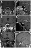
Figure 1
Figure 1
This 34-year-old woman presented with a 2-year history of galactorrhea and hyperprolactinemia, treated successfully with cabergoline. During previous treatment for Crohn disease, gadolinium-enhanced MRI showed a normal-appearing pituitary gland with Rathke cleft cyst (RCC) in the intermediate lobe ((A), axial; (B), sagittal). At 2-year follow-up, she had progressive headaches and her menstrual periods had ceased. MRI demonstrated the RCC and definite enlargement of the pituitary ((C), axial; (D), sagittal). No definitive adenoma was identifiable. Endocrine labs demonstrated normal prolactin, low gonadotroph and estrogen levels (central hypogonadism), and low normal thyroid hormone (all consistent with partial hypopituitarism). She underwent pituitary biopsy and drainage of the RCC. Pathological evaluation demonstrated ciliated low cuboidal epithelium and surrounding chronic lymphocytic inflammation. The anterior pituitary gland had marked mononuclear inflammation. The diagnosis was non-necrotizing granulomatous hypophysitis. She was treated with 40 mg/0.8 mL adalimumab weekly. Her headaches resolved but she developed progressive panhypopituitarism requiring replacement therapy. MRI demonstrated progressive pituitary atrophy 2 years later ((E), axial; (F), sagittal).
Figure 2
Figure 2
Coronal (A) and sagittal (B) T1-weighted MRI of a patient who initially presented with diabetes insipidus, showing focal nodular thickening and enhancement of the pituitary infundibulum with absence of neurohypophysis T1 signal. Biopsy and pathology found T cell predominant mixed inflammatory infiltrate. The clinicoradiologic picture was consistent with lymphocytic hypophysitis.
Figure 3
Figure 3
Coronal (A) and sagittal (B) T1-weighted MRI of a patient being treated with pembrolizumab that began experiencing signs of pituitary apoplexy with headache and extreme fatigue. T1 hypointense lesion is visible in the posterior left aspect of the pituitary gland, which is in the region of the previous T1 hyperintense posterior pituitary bright spot. Pathological analysis showed fragments of anterior and posterior pituitary gland with marked mononuclear inflammation and CD3+ T cell infiltrate. Reprinted with permission from Ref. [45] Copyright 2021. John Wiley & Sons.

References

    1. El Sayed S.A., Fahmy M.W., Schwartz J. Physiology, Pituitary Gland. StatPearls; Treasure Island, FL, USA: 2022. - PubMed
    1. Joshi M.N., Whitelaw B.C., Carroll P.V. Mechanisms in endocrinology: Hypophysitis: Diagnosis and treatment. Eur. J. Endocrinol. 2018;179:R151–R163. doi: 10.1530/EJE-17-0009. - DOI - PubMed
    1. Langlois F., Varlamov E.V., Fleseriu M. Hypophysitis, the growing spectrum of a rare pituitary disease. J. Clin. Endocrinol. Metab. 2022;107:10–28. doi: 10.1210/clinem/dgab672. - DOI - PMC - PubMed
    1. Prete A., Salvatori R. Hypophysitis. In: Feingold K.R., Anawalt B., Boyce A., Chrousos G., de Herder W.W., Dhatariya K., Dungan K., Hershman J.M., Hofland J., Kalra S., et al., editors. Endotext. MDText.com, Inc.; South Dartmouth, MA, USA: 2000.
    1. Gubbi S., Hannah-Shmouni F., Verbalis J.G., Koch C.A. Hypophysitis: An update on the novel forms, diagnosis and management of disorders of pituitary inflammation. Best. Pract. Res. Clin. Endocrinol. Metab. 2019;33:101371. doi: 10.1016/j.beem.2019.101371. - DOI - PMC - PubMed

LinkOut - more resources