. 2023 Mar 21;24(6):5942.
doi: 10.3390/ijms24065942.

Inflammatory Skin Disease Causes Anxiety Symptoms Leading to an Irreversible Course

Affiliations

Inflammatory Skin Disease Causes Anxiety Symptoms Leading to an Irreversible Course

Shohei Iida et al. Int J Mol Sci. .

Abstract

Intense itching significantly reduces the quality of life, and atopic dermatitis is associated with psychiatric conditions, such as anxiety and depression. Psoriasis, another inflammatory skin disease, is often complicated by psychiatric symptoms, including depression; however, the pathogenesis of these mediating factors is poorly understood. This study used a spontaneous dermatitis mouse model (KCASP1Tg) and evaluated the psychiatric symptoms. We also used Janus kinase (JAK) inhibitors to manage the behaviors. Gene expression analysis and RT-PCR of the cerebral cortex of KCASP1Tg and wild-type (WT) mice were performed to examine differences in mRNA expression. KCASP1Tg mice had lower activity, higher anxiety-like behavior, and abnormal behavior. The mRNA expression of S100a8 and Lipocalin 2 (Lcn2) in the brain regions was higher in KCASP1Tg mice. Furthermore, IL-1β stimulation increased Lcn2 mRNA expression in astrocyte cultures. KCASP1Tg mice had predominantly elevated plasma Lcn2 compared to WT mice, which improved with JAK inhibition, but behavioral abnormalities in KCASP1Tg mice did not improve, despite JAK inhibition. In summary, our data revealed that Lcn2 is closely associated with anxiety symptoms, but the anxiety and depression symptoms caused by chronic skin inflammation may be irreversible. This study demonstrated that active control of skin inflammation is essential for preventing anxiety.

Keywords: JAK inhibitor; Lipocalin 2; S100a8; anxiety; atopic dermatitis; cytokine; inflammatory skin; mouse model; psoriasis.


Conflict of interest statement

The authors declare no conflict of interest. The funders had no role in the study design; collection, analyses, or interpretation of data; writing of the manuscript; or decision to publish the results.

Figures

Figure 1
Figure 1
Behavioral experiments and JAK inhibitor treatment schedule; improvement of skin eruptions with JAK Inhibitor administration. (a) Schematic diagram of experimental procedures. (b) In 18-week-old KCASP1Tg mice, cerdulatinib treatment improved facial, head, and neck skin eruption. In WT mice, cerdulatinib treatment did not significantly alter skin symptoms compared with PBS treatment. (c) 18-week-old KCASP1Tg mice had lower body and whole brain weight compared to WT mice. Head and neck skin eruption of the mice was also evaluated using other sensory tests, using the EASI score, which is mainly used for the evaluation of atopic dermatitis (full score 7.2). The EASI score of KCASP1Tg mice significantly improved after cerdulatinib treatment. All data are expressed as the mean ± SEM by standard one-way ANOVA test, followed by Tukey’s multiple comparison test (*** p < 0.001, **** p < 0.0001). GHNS, general health and neurological screen; LD, light/dark transition test; OF, open field test; EP, elevated plus maze test; HP, hot plate test; SI, social interaction test; RR, rotarod test; CSI, three-chamber test; PPI, prepulse inhibition test; PS, Porsolt forced swim test; TM, T-maze spontaneous alternation test; TS, tail suspension test; FZ, contextual and cued fear conditioning test.
Figure 2
Figure 2
Behavioral analysis of four groups of KCASP1Tg and WT mice treated with PBS or cerdulatinib, respectively. (a) General health and neurological screen; KCASP1Tg mice had decreased body weights compared to WT mice. The grip strength of KCASP1Tg mice was not significantly different from that of WT mice. (b) Light/dark transition test; KCASP1Tg mice covered less distance in the dark chamber than WT mice and showed a decrease in the number of movements between the light and dark chambers, suggesting lower activity and higher anxiety-like behavior. Still, there was no predominant difference in the time spent in the light chamber. (c) Open field test; KCASP1Tg mice traveled less distance and spent less time in the center than WT mice, suggesting lower activity and higher anxiety tendencies. Cerdulatinib treatment did not improve distance traveled or center time in KCASP1Tg mice. (d) Elevated plus maze test; KCASP1Tg mice showed a decrease in distance traveled and the number of entries into the open and enclosed arms compared to WT mice, which implied low activity. There was no significant difference in the percentage of time spent in open arms. No behavioral changes were observed in KCASP1Tg mice after treatment with cerdulatinib. (e) Hot plate test; there was no difference in the predominance of pain sensitivity in the hot plate test between KCASP1Tg and WT mice. (f) Social interaction test; KCASP1Tg mice showed a decrease in distance traveled compared to WT mice, but there was no significant difference in the number of contacts or contact duration. (g) Rotarod test; KCASP1Tg mice showed no differences in coordinated locomotion or motor learning compared to WT mice. (h) Three-chamber test; KCASP1Tg mice spent less time in the cage containing stranger 1 than WT mice, indicating a decreased social behavior. In contrast, no difference was observed between KCASP1Tg and WT mice time spent around the cage containing stranger 1 and the time spent around the cage now containing a novel, unfamiliar mouse (stranger 2). (i) Acoustic startle response/prepulse inhibition test; KCASP1Tg mice showed reduced startle amplitude compared to WT mice, but there were no significant differences in prepulse inhibition compared to WT mice, suggesting a reduced response to acoustic stimuli. (j) Porsolt forced swim test; KCASP1Tg mice showed a decrease in immobility time on the second day compared to WT mice, suggesting that they may be more panicked. (k) T-maze spontaneous alternation test; KCASP1Tg mice took longer than WT mice but were not significantly different in correct responses or distance traveled. This suggests no deficit in working memory in KCASP1Tg mice. (l) Tail suspension test; KCASP1Tg mice were more active when suspended by the tail than WT mice. (m) Contextual and cued fear conditioning test; mice were exposed thrice to white noise as a conditioned stimulus (CS) for 30 s (horizontal black bars), followed by foot shock as an unconditioned stimulus (US) for the last 2 s of the conditioned stimulus (vertical arrows) during the conditioning session. Shock sensitivity is assessed based on the distance traveled during and after exposure to the unconditioned stimulus; there was no significant difference in immobility between KCASP1Tg and WT mice, suggesting no difference in contextual fear memory. All data are expressed as the mean ± SEM by ordinary one-way or two-way ANOVA, followed by Tukey’s multiple comparison test (* p < 0.05, ** p < 0.01, *** p < 0.001, **** p < 0.0001, ns: not significant).
Figure 2
Figure 2
Behavioral analysis of four groups of KCASP1Tg and WT mice treated with PBS or cerdulatinib, respectively. (a) General health and neurological screen; KCASP1Tg mice had decreased body weights compared to WT mice. The grip strength of KCASP1Tg mice was not significantly different from that of WT mice. (b) Light/dark transition test; KCASP1Tg mice covered less distance in the dark chamber than WT mice and showed a decrease in the number of movements between the light and dark chambers, suggesting lower activity and higher anxiety-like behavior. Still, there was no predominant difference in the time spent in the light chamber. (c) Open field test; KCASP1Tg mice traveled less distance and spent less time in the center than WT mice, suggesting lower activity and higher anxiety tendencies. Cerdulatinib treatment did not improve distance traveled or center time in KCASP1Tg mice. (d) Elevated plus maze test; KCASP1Tg mice showed a decrease in distance traveled and the number of entries into the open and enclosed arms compared to WT mice, which implied low activity. There was no significant difference in the percentage of time spent in open arms. No behavioral changes were observed in KCASP1Tg mice after treatment with cerdulatinib. (e) Hot plate test; there was no difference in the predominance of pain sensitivity in the hot plate test between KCASP1Tg and WT mice. (f) Social interaction test; KCASP1Tg mice showed a decrease in distance traveled compared to WT mice, but there was no significant difference in the number of contacts or contact duration. (g) Rotarod test; KCASP1Tg mice showed no differences in coordinated locomotion or motor learning compared to WT mice. (h) Three-chamber test; KCASP1Tg mice spent less time in the cage containing stranger 1 than WT mice, indicating a decreased social behavior. In contrast, no difference was observed between KCASP1Tg and WT mice time spent around the cage containing stranger 1 and the time spent around the cage now containing a novel, unfamiliar mouse (stranger 2). (i) Acoustic startle response/prepulse inhibition test; KCASP1Tg mice showed reduced startle amplitude compared to WT mice, but there were no significant differences in prepulse inhibition compared to WT mice, suggesting a reduced response to acoustic stimuli. (j) Porsolt forced swim test; KCASP1Tg mice showed a decrease in immobility time on the second day compared to WT mice, suggesting that they may be more panicked. (k) T-maze spontaneous alternation test; KCASP1Tg mice took longer than WT mice but were not significantly different in correct responses or distance traveled. This suggests no deficit in working memory in KCASP1Tg mice. (l) Tail suspension test; KCASP1Tg mice were more active when suspended by the tail than WT mice. (m) Contextual and cued fear conditioning test; mice were exposed thrice to white noise as a conditioned stimulus (CS) for 30 s (horizontal black bars), followed by foot shock as an unconditioned stimulus (US) for the last 2 s of the conditioned stimulus (vertical arrows) during the conditioning session. Shock sensitivity is assessed based on the distance traveled during and after exposure to the unconditioned stimulus; there was no significant difference in immobility between KCASP1Tg and WT mice, suggesting no difference in contextual fear memory. All data are expressed as the mean ± SEM by ordinary one-way or two-way ANOVA, followed by Tukey’s multiple comparison test (* p < 0.05, ** p < 0.01, *** p < 0.001, **** p < 0.0001, ns: not significant).
Figure 3
Figure 3
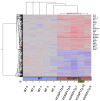
Microarray analysis of the cortex of 24-week-old KCASP1Tg and WT mice. Cerebral cortices of 24-week-old KCASP1Tg (n = 5) and WT (n = 5) mice were sampled for microarray analysis. Erdr1 (erythroid differentiation regulator 1) and Lcn2 (Neutrophil Gelatinase-associated Lipocalin; NGAL) gene expression was predominantly higher in KCASP1Tg mice compared to WT mice.
Figure 4
Figure 4
Lcn2 and S100a8 mRNA expression in the cortex, amygdala, hippocampus, and hypothalamus. After the completion of behavioral experiments, 18-week-old KCASP1Tg (n = 7) and WT (n = 10) mice were sacrificed, and each of the mRNA expressions of Lcn2 and S100a8, from four sites, were quantified using RT-PCR. Lcn2 and S100a8 were measured and standardized by the expression of GAPDH. KCASP1Tg mice had increased mRNA expression of Lcn2 and S100a8 in all regions of the cortex, amygdala, hippocampus, and hypothalamus compared to WT mice. In KCASP1Tg mice, cerdulatinib treatment decreased Lcn2 mRNA expression in the amygdala and hypothalamus, as well as S100a8 mRNA expression in the cortex, amygdala, and hypothalamus. All data were expressed as the mean ± SEM by standard one-way ANOVA test, followed by Tukey’s multiple comparison test (** p < 0.01, *** p < 0.001, **** p < 0.0001).
Figure 5
Figure 5
Lcn2 mRNA expression in astrocytes by inflammatory cytokine stimulation in vitro and Lcn2 in plasma. (a) We measured changes in mRNA expressions of the cultured astrocytes (n = 6) with inflammatory cytokines, including TNF-α, IL-17A, and IL-1β. Compared to other cytokines, stimulation with IL-1β increased Lcn2 mRNA expression in astrocytes. (b) At 18 weeks old, plasma Lcn2 was predominantly elevated in KCASP1Tg mice compared to WT mice. In KCASP1Tg mice, these decreased following cerdulatinib treatment. WT mice showed no significant difference after treatment with cerdulatinib. All data were expressed as the mean ± SEM by standard one-way ANOVA test, followed by Tukey’s multiple comparison test (*** p < 0.001, **** p < 0.0001).

Similar articles

Cited by

References

    1. Silverberg J.I. Comorbidities and the impact of atopic dermatitis. Ann. Allergy Asthma Immunol. 2019;123:144–151. doi: 10.1016/j.anai.2019.04.020. - DOI - PubMed
    1. Bakar R.S., Jaapar S.Z.S., Azmi A.F., Aun Y.C. Depression and anxiety among patients with psoriasis: A correlation with quality of life and associated factors. J. Taibah Univ. Med. Sci. 2021;16:491–496. doi: 10.1016/j.jtumed.2021.02.008. - DOI - PMC - PubMed
    1. Tiller J.W. Depression and anxiety. Med. J. Aust. 2013;199:S28–S31. doi: 10.5694/mja12.10628. - DOI - PubMed
    1. Mizutani H., Nishiguchi T., Murakami T. Animal Models of Atopic Dermatitis. Jpn. Med. Assoc. J. 2004;47:501–507. doi: 10.1097/01206501-200807000-00027. - DOI
    1. Kato S., Matsushima Y., Mizutani K., Kawakita F., Fujimoto M., Okada K., Kondo M., Habe K., Suzuki H., Mizutani H., et al. The Stenosis of Cerebral Arteries and Impaired Brain Glucose Uptake by Long-Lasting Inflammatory Cytokine Release from Dermatitis Is Rescued by Anti-IL-1 Therapy. J. Investig. Dermatol. 2018;138:2280–2283. doi: 10.1016/j.jid.2018.04.016. - DOI - PubMed

LinkOut - more resources