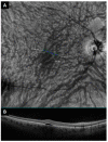
Intervortex Venous Anastomosis in the Macula in Central Serous Chorioretinopathy Imaged by En Face Optical Coherence Tomography
- PMID: 36983092
- PMCID: PMC10052017
- DOI: 10.3390/jcm12062088
Intervortex Venous Anastomosis in the Macula in Central Serous Chorioretinopathy Imaged by En Face Optical Coherence Tomography
Abstract
Purpose: To assess the presence of macular intervortex venous anastomosis in central serous chorioretinopathy (CSCR) patients using en face optical coherence tomography (EF-OCT).
Methods: A cross-sectional study where EF-OCT 6 × 6 and 12 × 12 mm macular scans of patients with unilateral chronic CSCR were evaluated for anastomosis between vortex vein systems in the central macula. The presence of prominent anastomoses was defined as a connection with a diameter ≥150 µm between the inferotemporal and superotemporal vortex vein systems which crossed the temporal raphe. Three groups were studied: CSCR eyes (with an active disease with the presence of neurosensorial detachment; n = 135), fellow unaffected eyes (n = 135), and healthy eyes as controls (n = 110). Asymmetries, abrupt termination, sausaging, bulbosities and corkscrew appearance were also assessed.
Results: In 79.2% of the CSCR eyes there were prominent anastomoses in the central macula between the inferotemporal and superotemporal vortex vein systems, being more frequent than in fellow eyes and controls (51.8% and 58.2% respectively). The number of anastomotic connections was higher in the affected eye group (2.9 ± 1.8) than in the unaffected fellow eye group (2.1 ± 1.7) and the controls (1.5 ± 1.6) (p < 0.001). Asymmetry, abrupt terminations and the corkscrew appearance of the choroidal vessels were more frequent in the affected eyes, although no differences in sausaging or bulbosities were observed.
Conclusions: Intervortex venous anastomoses in the macula were common in CSCR, being more frequently observed in affected eyes than in fellow unaffected eyes and healthy controls. This anatomical variation could have important implications concerning the pathogenesis and classification of the disease.
Keywords: central serous chorioretinopathy; choroidal vessels; en face optical coherence tomography; intervortex veins anastomosis; pachychoroid disease.
Conflict of interest statement
The authors declare that they have no conflict of interest.
Figures


References
-
- Spaide R.F., Rochepeau C., Kodjikian L., Garcia M.A., Coulon C., Burillon C., Denis P., Delaunay B., Mathis T., Matet A., et al. Choriocapillaris Flow Features Follow a Power Law Distribution: Implications for Characterization and Mechanisms of Disease Progression. Graefe’s Arch. Clin. Exp. Ophthalmol. 2019;257:905–912. doi: 10.1016/j.ajo.2016.07.023. - DOI - PubMed
LinkOut - more resources
Full Text Sources
Research Materials

