Review
. 2023 Mar 4;9(3):319.
doi: 10.3390/jof9030319.

Fusarium Photoreceptors

Affiliations
Review

Fusarium Photoreceptors

Javier Pardo-Medina et al. J Fungi (Basel). .

Abstract

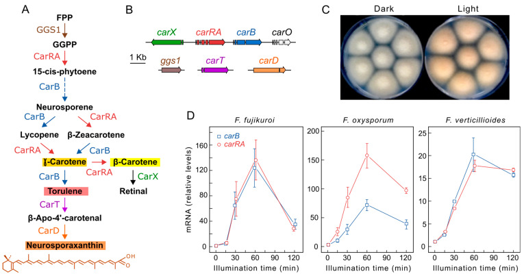
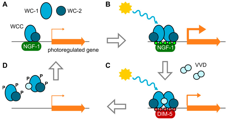
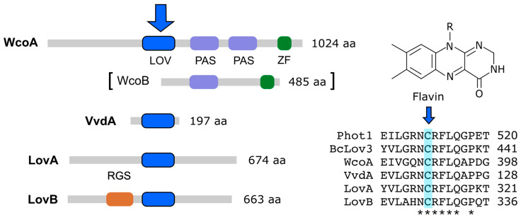
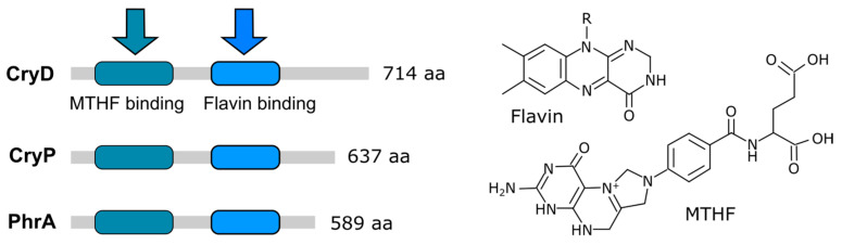
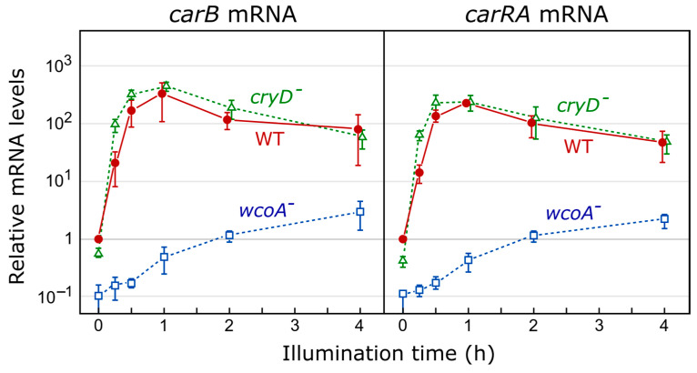
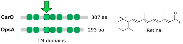
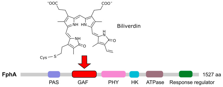
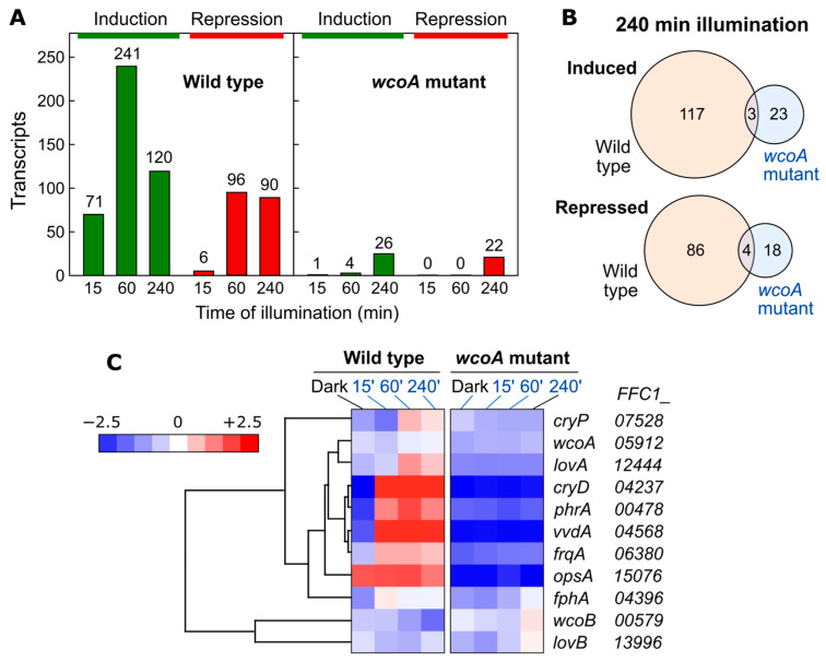
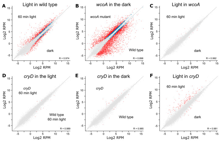
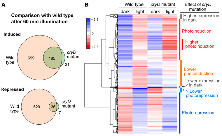
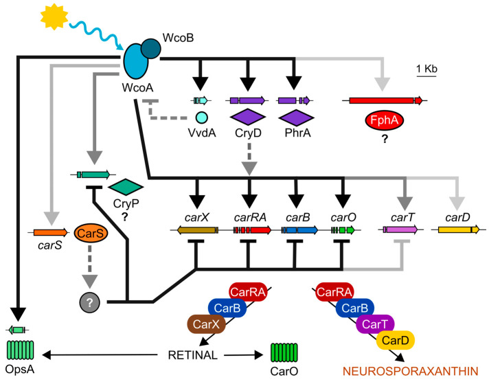
Light is an important modulating signal in fungi. Fusarium species stand out as research models for their phytopathogenic activity and their complex secondary metabolism. This includes the synthesis of carotenoids, whose induction by light is their best known photoregulated process. In these fungi, light also affects other metabolic pathways and developmental stages, such as the formation of conidia. Photoreceptor proteins are essential elements in signal transduction from light. Fusarium genomes contain genes for at least ten photoreceptors: four flavoproteins, one photolyase, two cryptochromes, two rhodopsins, and one phytochrome. Mutations in five of these genes provide information about their functions in light regulation, in which the flavoprotein WcoA, belonging to the White Collar (WC) family, plays a predominant role. Global transcriptomic techniques have opened new perspectives for the study of photoreceptor functions and have recently been used in Fusarium fujikuroi on a WC protein and a cryptochrome from the DASH family. The data showed that the WC protein participates in the transcriptional control of most of the photoregulated genes, as well as of many genes not regulated by light, while the DASH cryptochrome potentially plays a supporting role in the photoinduction of many genes.

Keywords: RNA-seq analyses; White Collar; cryptochrome; flavoprotein; light detection; phytochrome; rhodopsin.


Conflict of interest statement

The authors declare no conflict of interest. The funders had no role in the design of the study; in the collection, analyses, or interpretation of data; in the writing of the manuscript; in the decision to publish the results.

Figures

Figure 1
Figure 1
Photoregulation of carotenogenesis in Fusarium. (A) Carotenoid biosynthetic pathway. The colors of the main carotenoids that contribute to pigmentation in the mycelium are indicated in the background of their names. FPP: farnesyl diphosphate, GGPP: geranylgeranyl diphosphate. GGS1 is a prenyl transferase, CarRA is a bifunctional phytoene synthase/carotene cyclase, CarB is a desaturase, CarT and CarX are cleaving dioxygenases, CarD is an aldehyde dehydrogenase. (B) Genomic organization of the indicated genes in the pathway. The carO gene encodes a rhodopsin using retinal as chromophore. (C) Colonies of F. fujikuroi IMI58289 grown for one week in the dark or under illumination at 30 °C on minimal medium. (D) Light inductions of mRNA levels of the carRA and carB genes in three Fusarium species. Data from left to right adapted from [50] and from [42,44] (Copyright Elsevier, 2012 and 2016), respectively.
Figure 2
Figure 2
Simplified scheme of the mode of action of the White Collar complex (WCC) in N. crassa. (A) The WCC, consisting of WC-1 and WC-2, binds to the promoters of light-regulated genes in the dark. (B) Upon receipt of blue light, the interaction between WC-1 proteins forms WCC dimers that activate transcription through chromatin modifications (H3K14ac, small green circles) by histone acetyltransferase NGF-1. The transcription of many genes, including vvd, frq, and sub-1, is stimulated. (C) VVD protein accumulates in dimers in the nucleus and interacts with WC-1 in the WCC. New histone modification (H3K9me3, small red circles) by DIM-5 further contributes to stopping WCC activation. (D) WCCs dissociate from promoters and are transiently phosphorylated through PKC activity (not shown). WC-1 is partially degraded. After dephosphorylation or synthesis of new WC proteins in the dark, the cycle restarts. Adapted with permission from [1]; copyright 2016 American Society for Microbiology.
Figure 3
Figure 3
Flavin photoreceptors in Fusarium proteomes. Protein sizes and the major protein domains in F. fujikuroi are shown. The big blue arrow indicates the domain in which the chromophore, depicted on the right, is bound. WcoB is involved, as a partner of WcoA, in the WC complex. The LOV domains in LovA and LovB were identified through the NCBI domain search tool. Right: alignment of a 15-aa segment in the LOV domain of Phot1 from Arabidopsis thaliana (At3g45780), BcLov3 from B. cinerea (Bcin10g03870.1), and F. fujikuroi photoreceptors WcoA, VvdA, LovA, and LovB (Table 1). The conserved flavin-binding cysteine residue is shaded in blue and pointed with a small arrow. Numbers indicate the position of the last residue of the displayed segment in the corresponding protein.
Figure 4
Figure 4
Photoreceptors of the cryptochrome/photolyase family in Fusarium. Protein sizes and chromophore binding domains in F. fujikuroi are shown. The arrows indicate the binding of the chromophores that are depicted on the right. MTHF: 5,10-methenyltetrahydrofolate.
Figure 5
Figure 5
Effect of wcoA or cryD mutations on light induction of mRNA levels of the carRA and carB genes in F. fujikuroi. Cultures grown in shake flasks in the dark were transferred to Petri dishes, adapted to these conditions for 4 h, and then illuminated for the indicated intervals. mRNA levels are referred to those of the wild type (WT) in the dark. Adapted from [137,138].
Figure 6
Figure 6
Rhodopsins in Fusarium proteomes. Protein sizes and transmembrane domains in F. fujikuroi rhodopsins are shown. The two proteins bind to the retinal chromophore. The arrow indicates the transmembranal (TM) domain in which the chromophore is covalently attached to a conserved lysine residue.
Figure 7
Figure 7
Chromophore and domain structure of the Fusarium phytochrome. The F. fujikuroi coding gene fphA (FFUJ_05887) was identified due to its sequence similarity with F. graminearum Fgfph (FGSG_08608), which has been previously described [32]. The arrow indicates the domain in which the chromophore is expected to bind to a conserved cysteine residue.
Figure 8
Figure 8
Effect of illumination on wild-type (FKMC1995) and wcoA mutant transcriptomes. (A) Numbers of genes upregulated (green) or downregulated (red) after 15, 60, or 240 min of illumination using a four-fold threshold. (B) Venn diagrams of genes induced (above) or repressed (below) after 240 min illumination in the wild type and wcoA mutant. The intersection shows the coincident genes in both strains. (C) Hierarchical heatmap for the effect of different illumination intervals on transcript levels for photoreceptor genes in wild type and wcoA mutant. The FFC1_ numbers of the genes in the genome annotation are indicated on the right. The N. crassa frequency protein orthologous gene (frqA) is also included because of its connection to light regulation. Data adapted from [138].
Figure 9
Figure 9
Scatter plot representations of the effect of wcoA or cryD mutations on the transcriptomic response under 60 min illumination. Differentially expressed genes according to the DESeq2 analysis of the Seqmonk program are indicated in blue. Genes that exceed log2 values of ±2 are indicated in red. (A) Effect of light on the wild type. (B) Effect of wcoA mutation in the dark. (C) Effect of light on the wcoA mutant. (D) Effect of cryD mutation after 60 min of illumination. (E) Effect of cryD mutation in the dark. (F) Effect of light on the cryD mutant. Data from [137,138].
Figure 10
Figure 10
Effect of illumination on wild-type and cryD mutant transcriptomes. (A) Venn diagrams of the genes induced (above) or repressed (below) after 60 min illumination in the wild-type strain and in the cryD mutant (SG237) using a two-fold threshold. The intersection shows the coincident genes in both strains. (B) Hierarchical heatmap of genes induced or repressed after 60 min of illumination in the wild strain or in the cryD mutant. On the right are the groups, shown according to the effect of the cryD mutation. Data from [137].
Figure 11
Figure 11
Summary of the regulatory relationships between photoreceptors and carotenogenic genes in F. fujikuroi according to transcriptomic and experimental data from previous works. The intensity of the arrows represents the impact of each element on their targets. CarS is a RING-finger protein that downregulates the carotenoid pathway. Dashed lines indicate post-transcriptional effects. The grey element corresponds to a hypothetical regulator that is not yet known.

Similar articles

Cited by

References

    1. Fischer R., Aguirre J., Herrera-Estrella A., Corrochano L.M. The Complexity of Fungal Vision. Microbiol. Spectr. 2016;4:1–22. doi: 10.1128/microbiolspec.FUNK-0020-2016. - DOI - PubMed
    1. Tisch D., Schmoll M. Light Regulation of Metabolic Pathways in Fungi. Appl. Microbiol. Biotechnol. 2010;85:1259–1277. doi: 10.1007/s00253-009-2320-1. - DOI - PMC - PubMed
    1. Yu Z., Fischer R. Light Sensing and Responses in Fungi. Nat. Rev. Microbiol. 2019;17:25–36. doi: 10.1038/s41579-018-0109-x. - DOI - PubMed
    1. Chen C.-H., Ringelberg C.S., Gross R.H., Dunlap J.C., Loros J.J. Genome-Wide Analysis of Light-Inducible Responses Reveals Hierarchical Light Signalling in Neurospora. EMBO J. 2009;28:1029–1042. doi: 10.1038/emboj.2009.54. - DOI - PMC - PubMed
    1. Ruger-Herreros C., Rodríguez-Romero J., Fernández-Barranco R., Olmedo M., Fischer R., Corrochano L.M., Canovas D. Regulation of Conidiation by Light in Aspergillus nidulans. Genetics. 2011;188:809–822. doi: 10.1534/genetics.111.130096. - DOI - PMC - PubMed