Case Reports
. 2023 Feb 21;13(3):597.
doi: 10.3390/life13030597.

Duodenal Gangliocytic Paragangliomas-Case Series and Literature Review

Affiliations
Case Reports

Duodenal Gangliocytic Paragangliomas-Case Series and Literature Review

Madalina Stan-Ilie et al. Life (Basel). .

Abstract

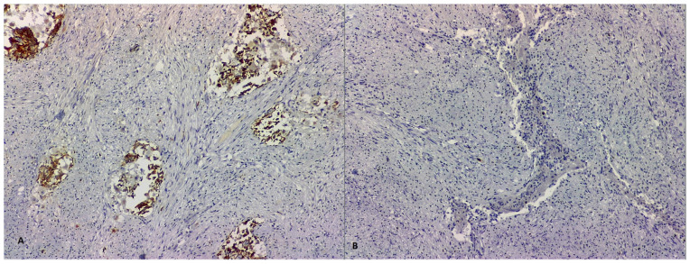
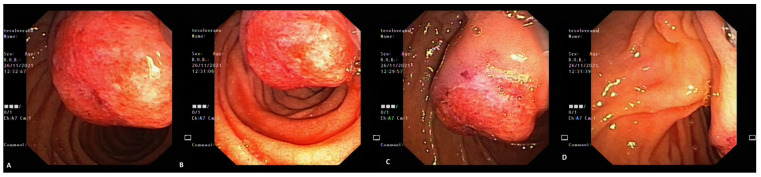
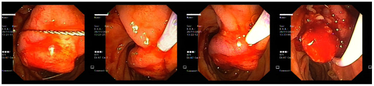
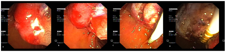
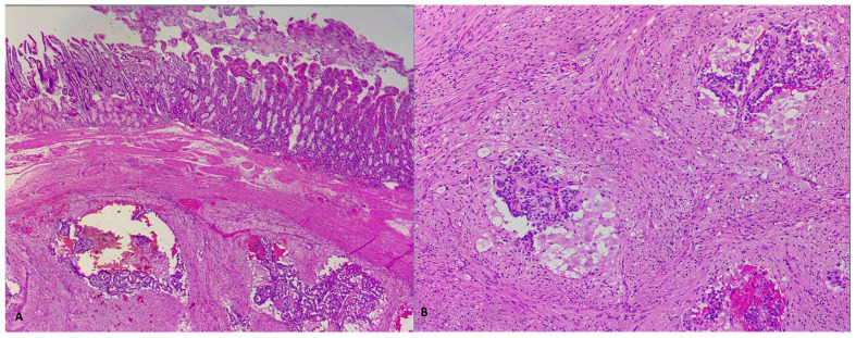
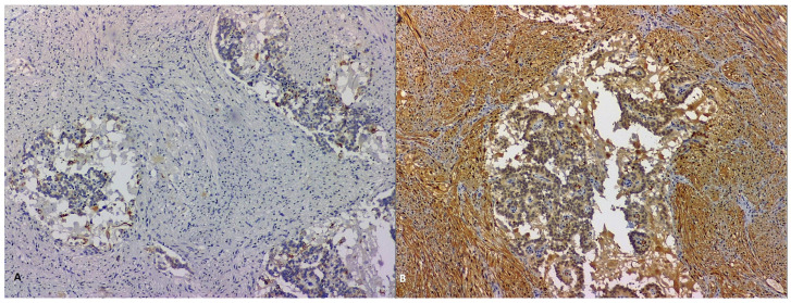
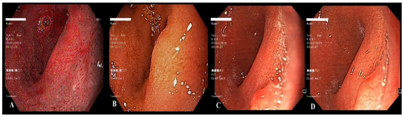
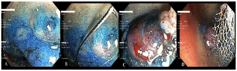
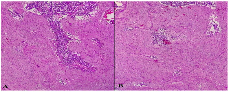
Duodenal gangliocytic paragangliomas are rare neuroendocrine tumors primarily localized in the periampullary area. Though mostly asymptomatic, they can present with various symptoms, most often jaundice, anemia and abdominal pain. The present paper is a case series report, describing our personal experience with patients presenting to the Emergency Unit with different symptoms due to duodenal gangliocytic paraganglioma. Endoscopic resection is safe and indicated in most of the cases, being also associated with lower medical costs. EUS plays a central role in the pre-resection management and in surveillance, and immunostaining is decisive to ascertain the tumor histologic origin. In addition to reporting our experience, we researched the literature regarding these rare tumors and performed a comprehensive review.

Keywords: duodenal gangliocytic paraganglioma; endoscopy; gastrointestinal bleeding; immunohistochemistry; jaundice; surgery.


Conflict of interest statement

The authors declare no conflict of interest.

Figures

Figure 1
Figure 1
ERCP image of the calculus identified by EUS and of the distal common bile duct stenosis (A) through which a plastic stent was inserted (B).
Figure 2
Figure 2
(A) EUS image revealing a submucosal lesion in the second part of the duodenum, mixed at elastography (arrow-mixed pattern of the lesion). (B) EUS guided-biopsy through EUS-FNB (arrow-needle inside the lesion).
Figure 3
Figure 3
CT (A) and MRI (B) aspects of the patient in the presurgical step. Arrow—duodenal location of the tumor.
Figure 4
Figure 4
HE-stained examination of tumoral proliferation typical of GP, showing fusiform, epithelial and ganglionar cells (A,B, 400×).
Figure 5
Figure 5
Microscopic immunohistochemical analysis showing chromogranin-positive staining of epithelial cells, but negative staining of fusiform and ganglionar cells (A, 100×), along with very rare, Ki67-positive tumoral cells (B, 100×).
Figure 6
Figure 6
Tumoral inspection with the side-viewing duodenoscope (AC) and bile flowing through the papilla right next to the tumor (D).
Figure 7
Figure 7
Endoscopic view during tumoral resection.
Figure 8
Figure 8
Endoscopic mixed hemostasis for the endoscopic resection site: injection of adrenaline 1:10,000 (A,B) and bipolar hemostasis (C,D).
Figure 9
Figure 9
Morphological and HE-stained samples to examine tumoral proliferation typical of GP covered by normal duodenal layers (A, 50×) and showing fusiform, epithelial and ganglionar cells (B, 200×).
Figure 10
Figure 10
Immunohistochemical analysis of the identified GP tumor showing pan-cytokeratin-focally positive epithelial cells (A, 100×) and S100-positive fusiform cells (B, 100×).
Figure 11
Figure 11
Polypoid lesion in the bulbar portion of the duodenum, right behind the pyloric sphincterial wall, inapparent at first glance (A,B), identified after a complete endoscopic exam (C,D).
Figure 12
Figure 12
Examination by a lateral-viewing duodenoscope (A), electroexcision (B,C) and retrieval of the polyp (D).
Figure 13
Figure 13
Morphological and standard coloration aspects of the tumor showing large ganglionar tumor cell proliferation (A, 100×) and fusiform cells (B, 100×).
Figure 14
Figure 14
PET whole-body scanning showing the absence of tumoral relapse.

References

    1. Bakshi N., Rao S., Dhawan S., Singla V. Duodenal gangliocytic paraganglioma: A rare cause for gastrointestinal polyp. Indian J. Pathol. Microbiol. 2018;61:580–582. doi: 10.4103/IJPM.IJPM_479_17. - DOI - PubMed
    1. Okubo Y., Yokose T., Motohashi O., Miyagi Y., Yoshioka E., Suzuki M., Washimi K., Kawachi K., Nito M., Nemoto T., et al. Duodenal Rare Neuroendocrine Tumor: Clinicopathological Characteristics of Patients with Gangliocytic Paraganglioma. Gastroenterol. Res. Pract. 2016;2016:5257312. doi: 10.1155/2016/5257312. - DOI - PMC - PubMed
    1. Sharma S., Gaspar B.L., Kumar P., Yadav T.D., Vasishta R.K. Gangliocytic paraganglioma with atypical immunohistochemical features presenting as extrahepatic biliary obstruction. Int. J. Surg. Pathol. 2015;23:561–566. doi: 10.1177/1066896915589969. - DOI - PubMed
    1. Park H.K., Han H.S. Duodenal Gangliocytic Paraganglioma With Lymph Node Metastasis. Arch. Pathol. Lab. Med. 2016;140:94–98. doi: 10.5858/arpa.2014-0456-RS. - DOI - PubMed
    1. Riley D.S., Barber M.S., Kienle G.S., Aronson J.K., von Schoen-Angerer T., Tugwell P., Kiene H., Helfand M., Altman D.G., Sox H., et al. CARE guidelines for case reports: Explanation and elaboration document. J. Clin. Epi. 2017;89:218–235. doi: 10.1016/j.jclinepi.2017.04.026. - DOI - PubMed

Publication types

LinkOut - more resources