Review
. 2023 Mar 17;13(3):818.
doi: 10.3390/life13030818.

Metastasis in Neuroblastoma and Its Link to Autophagy

Affiliations
Review

Metastasis in Neuroblastoma and Its Link to Autophagy

Leila Jahangiri. Life (Basel). .

Abstract

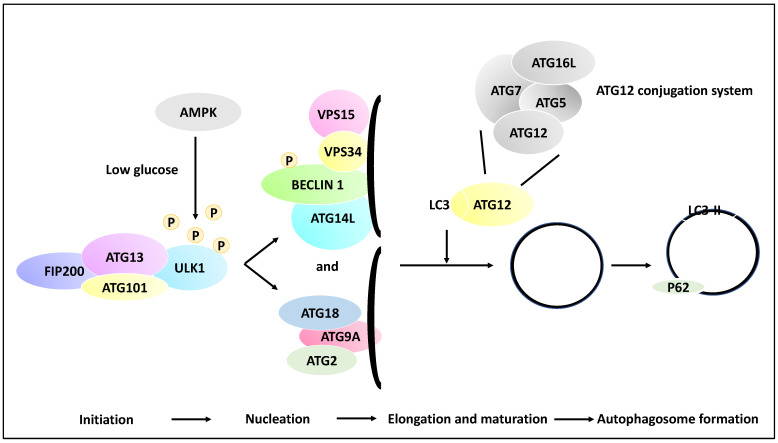
Neuroblastoma is a paediatric malignancy originating from the neural crest that commonly occurs in the abdomen and adrenal gland, leading to cancer-related deaths in children. Distant metastasis can be encountered at diagnosis in greater than half of these neuroblastoma patients. Autophagy, a self-degradative process, plays a key role in stress-related responses and the survival of cells and has been studied in neuroblastoma. Accordingly, in the early stages of metastasis, autophagy may suppress cancer cell invasion and migration, while its role may be reversed in later stages, and it may facilitate metastasis by enhancing cancer cell survival. To that end, a body of literature has revealed the mechanistic link between autophagy and metastasis in neuroblastoma in multiple steps of the metastatic cascade, including cancer cell invasion and migration, anoikis resistance, cancer cell dormancy, micrometastasis, and metastatic outbreak. This review aims to take a step forward and discuss the significance of multiple molecular players and compounds that may link autophagy to metastasis and map their function to various metastatic steps in neuroblastoma.

Keywords: autophagy; metastasis; neuroblastoma; paediatric cancers.


Conflict of interest statement

The author declares no conflict of interest.

Figures

Figure 1
Figure 1
The roles of autophagy in metastasis. (A) The primary tumour cells (for example, neuroblastoma cells) during the in-situ step may be affected by autophagy; for example, autophagy may inhibit metastasis by limiting pro-metastasis inflammatory responses and limiting growth (−), or it can promote metastasis by increasing drug resistance (+). (B) Tumour cells (for example, neuroblastoma cells) can enter the circulation, constituting circulating tumour cells, and autophagy promotes resistance to anoikis (anoikis is triggered by a contact loss with the basement membrane), hence promoting metastasis (+). Arrowhead showing tumour cells entering the circulation. (C) At the extravasation and seeding step, the circulating tumour cells enter a distant site, and autophagy may trigger the onset of dormancy in the upper panel of (C) (the cell with a slightly darker yellow colour represents a dormant neuroblastoma cell) or survival in the new microenvironment (+) and drug resistance (+) in the bottom panel of (C). Arrowhead showing tumour cells exiting the circulation. (D) In the distant metastasis site, autophagy can limit the expansion of dormant tumour cells (+/−) in the upper panel of (D) or promote adaptation to this new environment, expansion and metastatic outbreak (+) in the bottom panel of (D). It is noteworthy that (+) and (−) signs in the text signify a positive or negative contribution of autophagy to metastasis, respectively, while the grey basement layer in (A,B) and pink basement layer in (C,D) represent distinct tumour microenvironments before and after migration, respectively.
Figure 2
Figure 2
Multiple molecular steps of autophagy. A pre-autophagosomal structure is formed in the initiation stage and comprises ULK1, FIP200, ATG101 and ATG13, and in low glucose states, the ULK1 may receive activating phosphorylation via AMPK activity (whereas in the high-glucose state, ULK1 receives inhibitory phosphorylation due to mTOR and raptor activity, although this is not shown in the figure). ULK1 then activates the nucleation of the phagophore and the phosphorylation of Beclin 1. In the nucleation phase, ATG9A is recruited to the isolation membrane (phagophore), and the recruitment of ATG2 and ATG18 ensues. In addition, the isolation membrane is coated with VPS34, VPS15, ATG14L, and Beclin 1. Further, in the elongation and maturation phase, the processing of LC3 and ATG12 conjugation systems leads to the elongation of the phagophore and the formation of autophagosomes. It is noteworthy that the ATG12 conjugation system comprises ATG12, ATG7, ATG16L and ATG5 proteins. These processes contribute to the coating of the autophagosome with proteins such as LC3-II and P62, while the formed autophagosome may fuse with a lysosome in later stages.
Figure 3
Figure 3
The contribution of ULK1 to NB apoptosis and metastasis. (A) ULK1 forms complexes with ATG13, ATG101 and FIP200. ULK1 can be activated by AMPK via activating phosphorylation when glucose levels are low. ULK1 then can contribute to the activation of nucleation of the phagophore and the phosphorylation of Beclin 1. (B) The addition of SBI-0206965, a small molecule inhibitor of ULK1, led to the inhibition of autophagy marked by an increase in P62, a decrease in ULK1, LC3 lipidation, and LC3-II levels in the presence of bafilomycin A1. Therefore, SBI-0206965 decreased autophagic flux. (C) SBI-0206965 treatment in NB cell lines led to the upregulation of PARP and caspase-3, while cell viability was decreased, suggesting that the inhibition of ULK1 could lead to anoikis (hence ULK1 leads to anoikis resistance and perhaps metastasis). (D) The genetic inhibition model, SK-N-AS cells expressing a dominant-negative ULK1 gene (e.g., dnULK1K46N), similar to SBI-0206965, led to increased cleaved PARP, annexin-V, caspase-3 and enhanced enzymatic activity of caspase-3/7 and 8 and may also promote anoikis (hence the presence of ULK1 can lead to anoikis resistance). (E) Xenografting of SK-N-AS cells expressing stable dnULK1 led to P62 accumulation, increased PARP and caspase-3 levels, and a reduced metastasis burden in the liver in this group compared to their control counterparts. (F) TRAIL and SBI-0206965 combination in SK-N-AS cells led to increased apoptosis. TRAIL treatment, per se, increased autophagic flux, suggesting that perhaps autophagy has been upregulated to counter TRAIL-mediated apoptosis levels.
Figure 4
Figure 4
NORAD-mediated NB progression. (A) The knockdown of lncRNA NORAD using siRNAs reduces proliferation, invasion, and DOX resistance in NB cell lines, including SK-N-SH and IMR-32. In addition, NORAD knockdown may lead to P62 downregulation and increased levels of Beclin 1, ATG5, LC3-II/LC3-I (LC3-II/1), and enhanced apoptosis. (B) miR-144-3p was a target of NORAD, depicted by the binding of NORAD (blue) to miR-144-3p (red). (C) Further, the overexpression of NORAD led to the downregulation of miR-144-3p, resulting in cancer progression and DOX resistance. (D) The depletion of miR-144-3p (red) using anti-miR-144-3p (purple) reduced the suppressive effects of siRNA-mediated NORAD depletion on NB cell proliferation, migration, metastasis, and DOX resistance (NORAD and NORAD siRNA have been depicted in blue and black colours, respectively). (E) Histone deacetylase 8 (HDAC8) was shown to be a target of miR-144-3p in NB. miR-144-3p expression (accumulation) led to the downregulation of protein levels of HDAC8, while anti-miR-144-3p treatment led to HDAC8 upregulation. In addition, NORAD depletion led to reduced HDAC8 expression, collectively suggesting HDAC8 was regulated through the NORAD/miR-144-3p axis. (F) NORAD depletion in a murine xenograft model led to reduced tumour growth.
Figure 5
Figure 5
The role of MEG3 in regulating autophagy in NB metastasis. (A) MEG3, a nucleus-based lncRNA, was tested in NB cell lines, including SK-N-BE2, SK-N-AS, and SH-SY5Y. The overexpression of MEG3 led to reduced cell proliferation, colony formation, migration, and invasion capacity. In addition, MEG3 overexpression reduced autophagy protein levels, including ATG3, ATG12, and Beclin 1, but this effect was not implemented through mTORC1 since in MEG3-overexpressing NB cells, mTORC1 was inactivated. (B) A FOXO1 inhibitor (AS1842856) mimicked MEG3 overexpression and led to reduced LC3 protein levels (suppression of autophagy), while the overexpression of MEG3 suppressed FOXO1, suggesting perhaps that MEG3 attenuated autophagy through FOXO1 regulation. MEG3 downregulation led to enhanced levels of ATG16, ATG3, and Beclin 1 (in an mTOR-independent manner), while low levels of MEG3 enhanced migration through mTOR signalling.
Figure 6
Figure 6
Isatin reduced the invasion and metastasis capacity of NB cells. (A) SH-SY5Y cells were treated with Isatin and were subjected to microarray analysis, revealing the differential expression of genes involved in redox activities, transcription, transport, metabolism, chemokine and mTOR signalling, and ribosome-related pathways. (B) Isatin treatment led to the reduced invasion and migration capacity of SH-SY5Y NB cells through the inhibition of phosphorylated-mTOR (p-mTOR) and the increase of phosphorylated-AMPK (p-AMPK) (since AMPK inhibits mTOR). (C) Isatin treatment led to the upregulation of LC3-II and Beclin1 and the downregulation of P62.
Figure 7
Figure 7
Apatinib reduced the invasion and metastasis capacity of NB cells. (A) NB cell lines inclusive of BE(2)-M17, IMR-32 and SH-SY5Y were treated with apatinib leading to reduced viability, colony formation, migration and proliferation of these cells while increasing apoptosis rate (reduced Bcl-2/Bax ratio). (B) Apatinib treatment led to cell cycle arrest (reduced cyclin D1 levels) while decreasing phosphorylated mTOR, AKT, and ERK (p-mTOR, p-AKT and p-ERK, respectively). (C) LC3-II/I and ATG5 protein levels were increased as a result of apatinib treatment, revealing autophagy activation.
Figure 8
Figure 8
Summary flowchart of the molecular players and compounds reviewed in this study linking NB metastasis to autophagy. ULK1 and SHNG16 induced autophagy, metastasis, and MEG3 and miR-34a suppressed autophagy and metastasis; NORAD and hsa_circ_0013401 suppressed autophagy but induced metastasis, and Isatin, Apatinib and Honokiol induced autophagy but suppressed metastasis. Finally, 3-MA and HCQ may be useful inhibitors of autophagy to improve the efficacy of therapy in NB (for example, the combination of 3-MA with sulforaphane or the combination of HCQ with vincristine, reduced viability and progression of NB, respectively) and clinical trials may be launched to further investigate these links. It is noteworthy that in this figure, the arrows pointing upwards and downwards signify induction or suppression, respectively.

Similar articles

Cited by

References

    1. Bosse K.R., Maris J.M. Advances in the translational genomics of neuroblastoma: From improving risk stratification and revealing novel biology to identifying actionable genomic alterations. Cancer. 2016;122:20–33. doi: 10.1002/cncr.29706. - DOI - PMC - PubMed
    1. Maris J.M., Hogarty M.D., Bagatell R., Cohn S.L. Neuroblastoma. Lancet. 2007;369:2106–2120. doi: 10.1016/S0140-6736(07)60983-0. - DOI - PubMed
    1. Cohn S.L., Pearson A.D.J., London W.B., Monclair T., Ambros P.F., Brodeur G.M., Faldum A., Hero B., Iehara T., Machin D., et al. The International Neuroblastoma Risk Group (INRG) classification system: An INRG Task Force report. J. Clin. Oncol. 2009;27:289–297. doi: 10.1200/JCO.2008.16.6785. - DOI - PMC - PubMed
    1. Brodeur G.M. Neuroblastoma: Biological insights into a clinical enigma. Nat. Rev. Cancer. 2003;3:203–216. doi: 10.1038/nrc1014. - DOI - PubMed
    1. Brodeur G.M., Pritchard J., Berthold F., Carlsen N.L., Castel V., Castelberry R.P., De Bernardi B., Evans A.E., Favrot M., Hedborg F. Revisions of the international criteria for neuroblastoma diagnosis, staging, and response to treatment. J. Clin. Oncol. 1993;11:1466–1477. doi: 10.1200/JCO.1993.11.8.1466. - DOI - PubMed

LinkOut - more resources