. 2023 Mar 20;13(3):836.
doi: 10.3390/life13030836.

Detection of Lymphatic Vessels in the Superficial Fascia of the Abdomen

Affiliations

Detection of Lymphatic Vessels in the Superficial Fascia of the Abdomen

Giovanna Albertin et al. Life (Basel). .

Abstract

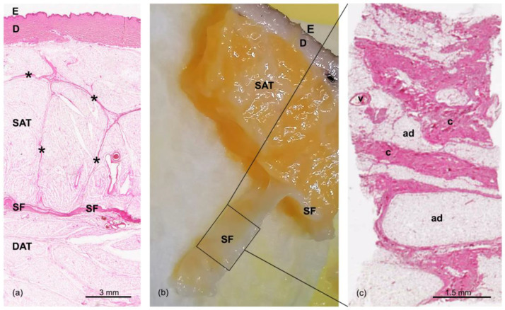
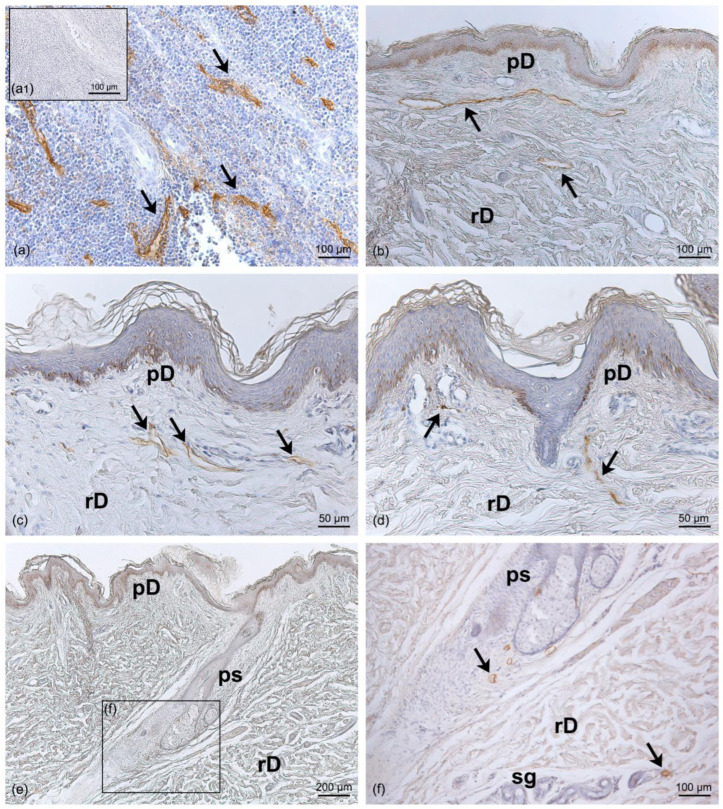
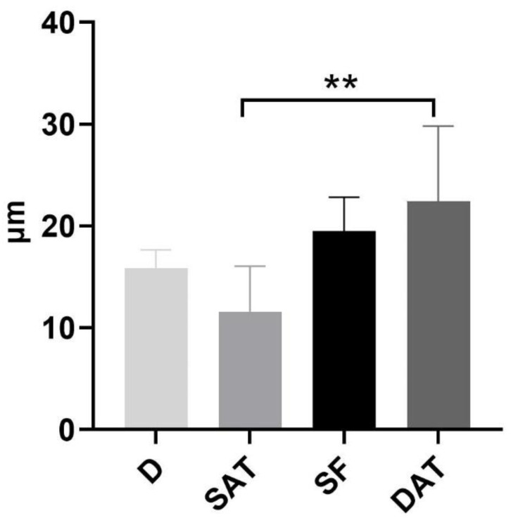
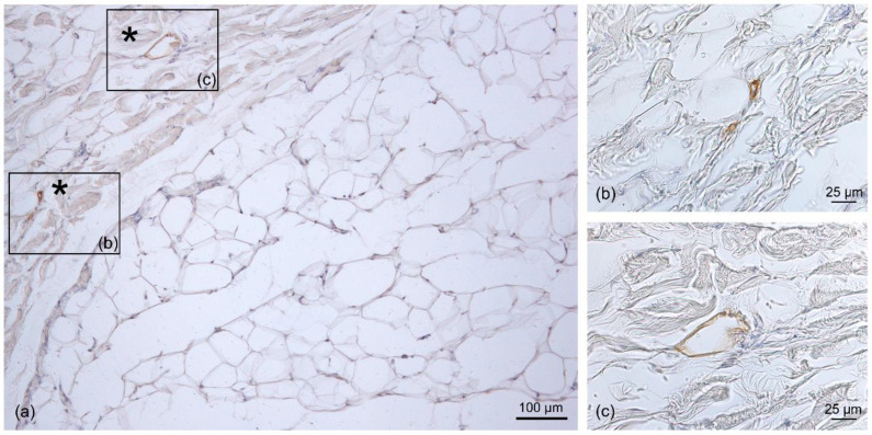
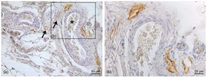
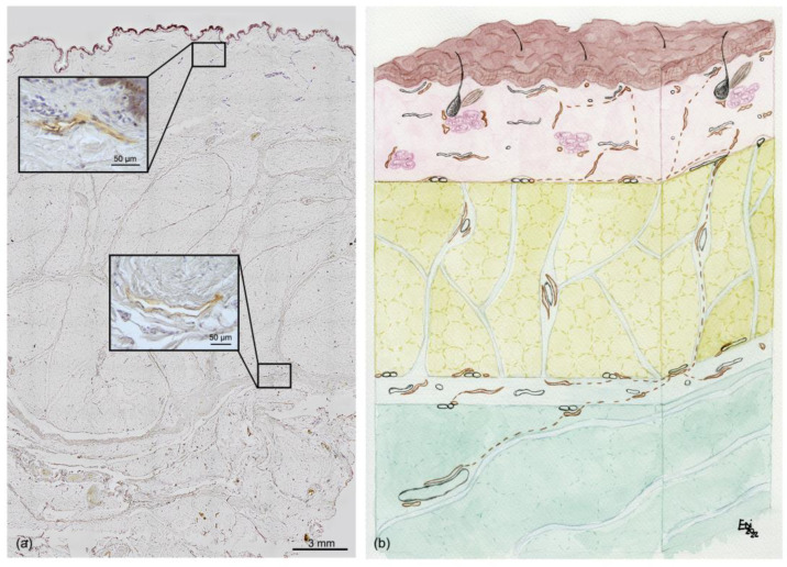
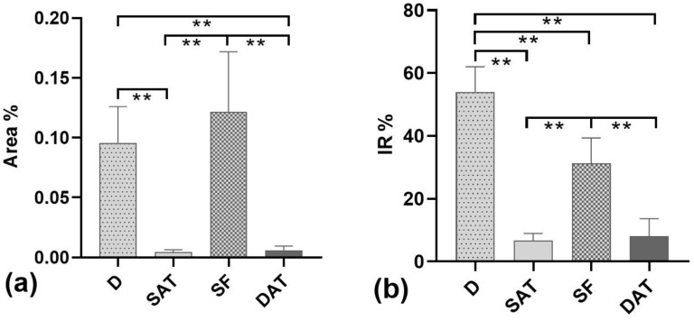
Recently, the superficial fascia has been recognized as a specific anatomical structure between the two adipose layers-the superficial adipose tissue (SAT) and the deep adipose tissue (DAT). The evaluation of specific characteristics of cells, fibers, blood circulation, and innervation has shown that the superficial fascia has a clear and distinct anatomical identity, but knowledge about lymphatic vessels in relation to the superficial fascia has not been described. The aim of this study was to evaluate the presence of lymphatic vessels in the hypodermis, with a specific focus on the superficial fascia and in relation to the layered subdivision of the subcutaneous tissue into SAT and DAT. Tissue specimens were harvested from three adult volunteer patients during abdominoplasty and stained with D2-40 antibody for the lymphatic endothelium. In the papillary dermis, a huge presence of lymphatic vessels was highlighted, parallel to the skin surface and embedded in the loose connective tissue. In the superficial adipose tissue, thin lymphatic vessels (mean diameter of 11.6 ± 7.71 µm) were found, close to the fibrous septa connecting the dermis to the deeper layers. The deep adipose tissue showed a comparable overall content of lymphatic vessels with respect to the superficial layer; they followed the blood vessel and had a larger diameter. In the superficial fascia, the lymphatic vessels showed higher density and a larger diameter, in both the longitudinal and transverse directions along the fibers, as well as vessels that intertwined with one another, forming a rich network of vessels. This study demonstrated a different distribution of the lymphatic vessels in the various subcutaneous layers, especially in the superficial fascia, and the demonstration of the variable gauge of the vessels leads us to believe that they play different functional roles in the collection and transport of interstitial fluid-important factors in various surgical and rehabilitation fields.

Keywords: hypodermis; immunohistochemistry; lymphatic system; lymphatics vessels; reconstructive surgery; subcutaneous tissue; superficial fascia.


Conflict of interest statement

The authors declare no conflict of interest.

Figures

Figure 1
Figure 1
Skin and the subcutaneous layers of the abdomen: (a) Hematoxylin and eosin staining of a full-thickness specimen, in which we can see the epidermis (E) and dermis (D); the superficial adipose tissue (SAT), organized in lobules separated by the fibrous septa of the retinacula cutis (asterisks); the superficial fascia (SF); and the deep adipose tissue (DAT). (b) Formalin-fixed sample of the abdominal region (E: epidermis, D: dermis; SAT: superficial adipose tissue, SF: superficial fascia). The SF—the layer localized between the SAT and DAT—was isolated and stained with hematoxylin and eosin. (c) A tangential section of the flat embedded SF shown in panel (b). The SF is formed by a network of collagen fibers arranged irregularly (c: connective tissue), interconnected and mixed with adipocytes (ad), and crossed by blood vessels (v). Scale bars: (a) = 3 mm; (b) = 1.5 mm.
Figure 2
Figure 2
Immunohistochemistry with D2-40 antibody on full-thickness specimens of the dermis of the abdominal region: (a) Positive control of immunoreaction for the D2-40 LV marker in human tonsil tissue, where the brown staining (arrows) highlights the LVs in the endothelium between the lymphoid tissues; (a1) inset of negative control with human tonsil tissue by omission of the D2-40 antibody. (bd) The distribution of LVs follows different orientations in the papillary dermis (pD) and the reticular dermis (rD) (arrows). (e) Full length of the pilosebaceous structure (ps) from the epidermis to the reticular dermis; the magnification in (f) shows LVs close to the pilosebaceous structure and sweat glands (sg) (black arrow). Scale bars: (a,b,a1,f) = 100 µm; (c,d) = 50 µm; (e) = 200 µm.
Figure 3
Figure 3
Mean diameter (µm) of LVs in specific areas—D, SAT, SF, and DAT. ** = Multiple comparison analyses found a significant difference in the vessel diameters between the SAT and the DAT (p = 0.038).
Figure 4
Figure 4
Immunohistochemistry with D2-40 antibody on a full-thickness-specimen of the SAT layer: (a) immunostaining of lymphatic vessels between collagen fibers (asterisk) at the fibrous septa level; (b,c) 40× magnification of the vessels. Scale bars: (a) = 100 µm; (b,c) = 25 µm.
Figure 5
Figure 5
Immunohistochemistry with D2-40 antibody on the superficial fascia (SF) layer: The LVs follow the collagen fibers (a) and are distributed close to the blood vessels (b). (c,d) Several sections of LVs (black arrow) and blood vessels (white arrows) between the collagen fibers (c) of the SF and the transverse section of the LVs (black asterisk) close to the transverse section of the blood vessels (white asterisk) (d); (c1) inset of the negative control with SF by omission of the D2-40 antibody. Scale bars: (a,c,c1,d) = 100 µm; (b) = 50 µm.
Figure 6
Figure 6
Immunohistochemistry with D2-40 antibody in a full-thickness specimen f the DAT layer: (a) Positive immunostaining of LVs (black arrow) close to the blood vessels (black asterisk); in the blood vessels, the endothelium is not stained, and large amounts of erythrocytes are visible in the lumen; (b) 20× Magnification: blood vessels with open LVs around them. Scale bars: (a) = 50 µm; (b) = 25 µm.
Figure 7
Figure 7
Immunohistochemistry and schematic drawing of the distribution of LVs in the subcutis: (a) Immunohistochemistry with D2-40 antibody on a full-thickness, with two high-magnification examples of staining with D2-40 (in the insets), on the dermis and superficial fascia, respectively (scale bar: 50 µm). (b) Schematic drawing of the distribution of LVs (rust color) in the skin and subcutaneous tissue. The LVs’ organization follows the layer organization. A first LV plexus is present at the dermis level up to the dermis border. Our results may indicate the presence of LVs with a vertical course following the retinacula cutis (dotted line) in the SAT; the vessels connect to form a second plexus in the superficial band and, even deeper, in the DAT, where the vessels go to form LVs of greater caliber (i.e., collectors) (scale bar: 200 µm).
Figure 8
Figure 8
Immunoreactivity % in the cutis and subcutis. (a) Percentage of the tissue area covered by immunoreactivity (Area%). (b) Percentages of immunoreactive structures (IR%) in the dermis (D), superficial adipose tissue (SAT), superficial fascia (SF), and deep adipose tissue (DAT); ** = p < 0.05 according to multiple comparison analyses.

References

    1. Oliver G., Kipnis J., Randolph G.J., Harvey N.L. The Lymphatic Vasculature in the 21st Century: Novel Functional Roles in Homeostasis and Disease. Cell. 2020;182:270–296. doi: 10.1016/j.cell.2020.06.039. - DOI - PMC - PubMed
    1. Weber E., Aglianò M., Bertelli E., Gabriele G., Gennaro P., Barone V. Lymphatic Collecting Vessels in Health and Disease: A Review of Histopathological Modifications in Lymphedema. Lymphat. Res. Biol. 2022;20:468–477. doi: 10.1089/lrb.2021.0090. - DOI - PMC - PubMed
    1. Mehrara B.J., Greene A.K. Lymphedema and obesity: Is there a link? Plast. Reconstr. Surg. 2014;134:154e–160e. doi: 10.1097/PRS.0000000000000268. - DOI - PMC - PubMed
    1. Schaverien M.V., Coroneos C.J. Surgical Treatment of Lymphedema. Plast. Reconstr. Surg. 2019;144:738–758. doi: 10.1097/PRS.0000000000005993. - DOI - PubMed
    1. Macchi V., Tiengo C., Porzionato A., Stecco C., Galli S., Vigato E., Azzena B., Parenti A., De Caro R. Anatomo-radiological study of the superficial musculo-aponeurotic system of the face. Ital. J. Anat. Embryol. 2007;112:247–253. - PubMed