Review
. 2023 Mar 21;59(3):622.
doi: 10.3390/medicina59030622.

Anatomical Variations of the External Jugular Vein: A Pictorial and Critical Review

Affiliations
Review

Anatomical Variations of the External Jugular Vein: A Pictorial and Critical Review

Mugurel Constantin Rusu et al. Medicina (Kaunas). .

Abstract

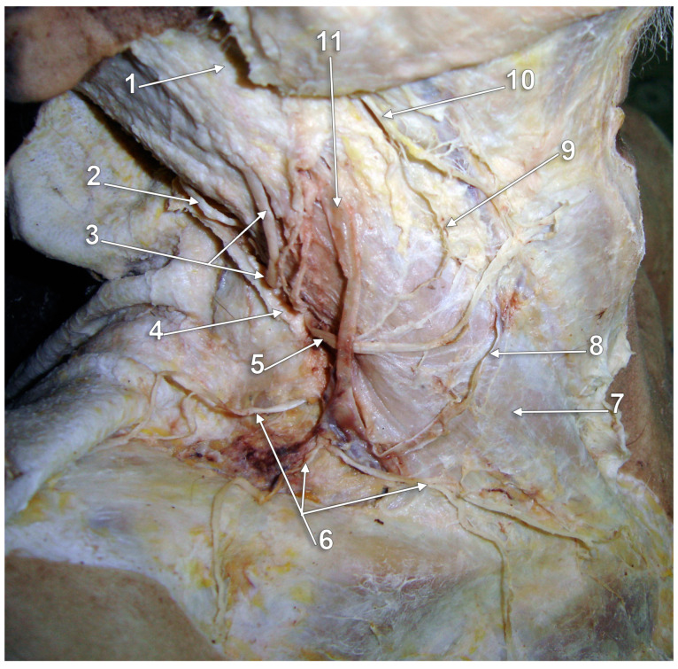
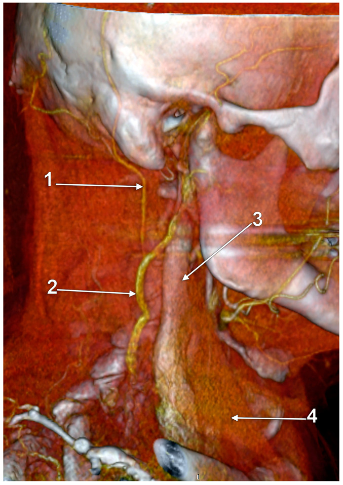
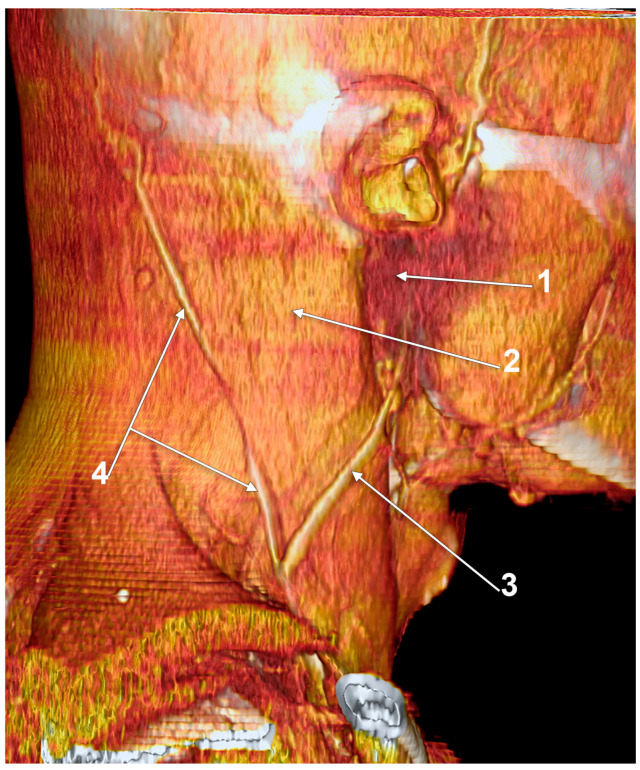
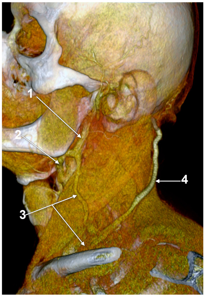
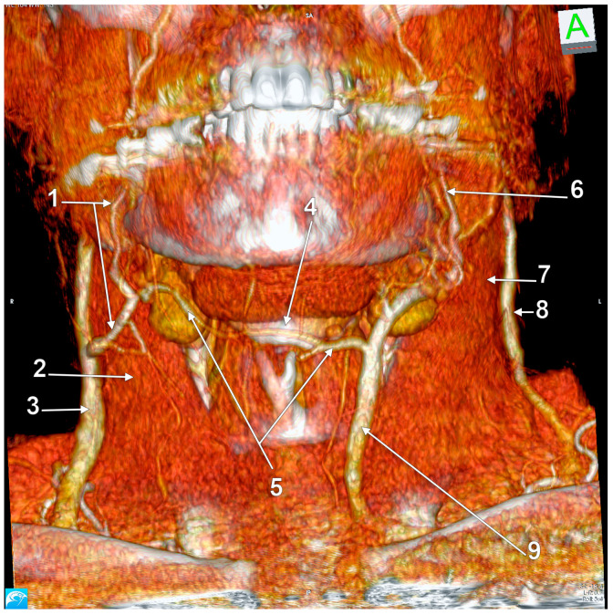
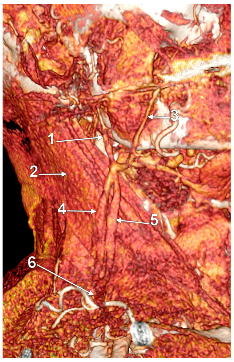
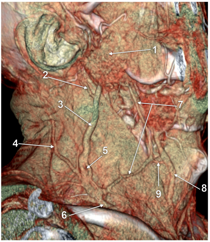
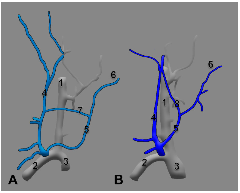
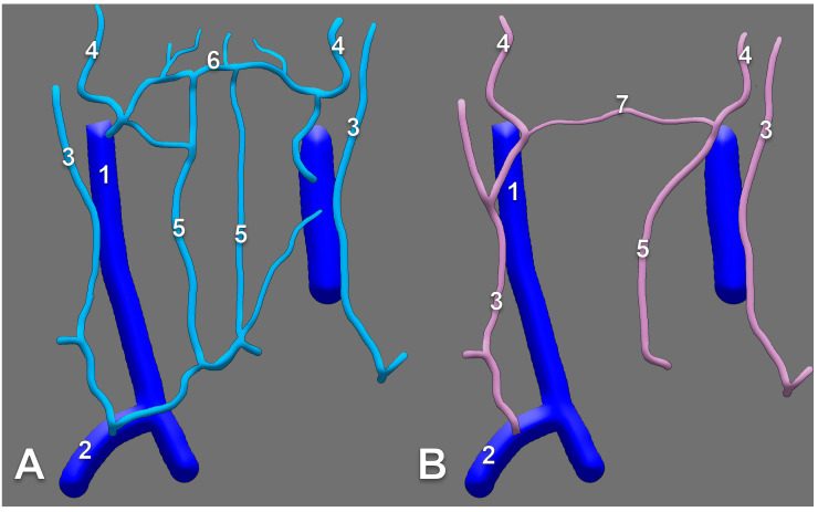
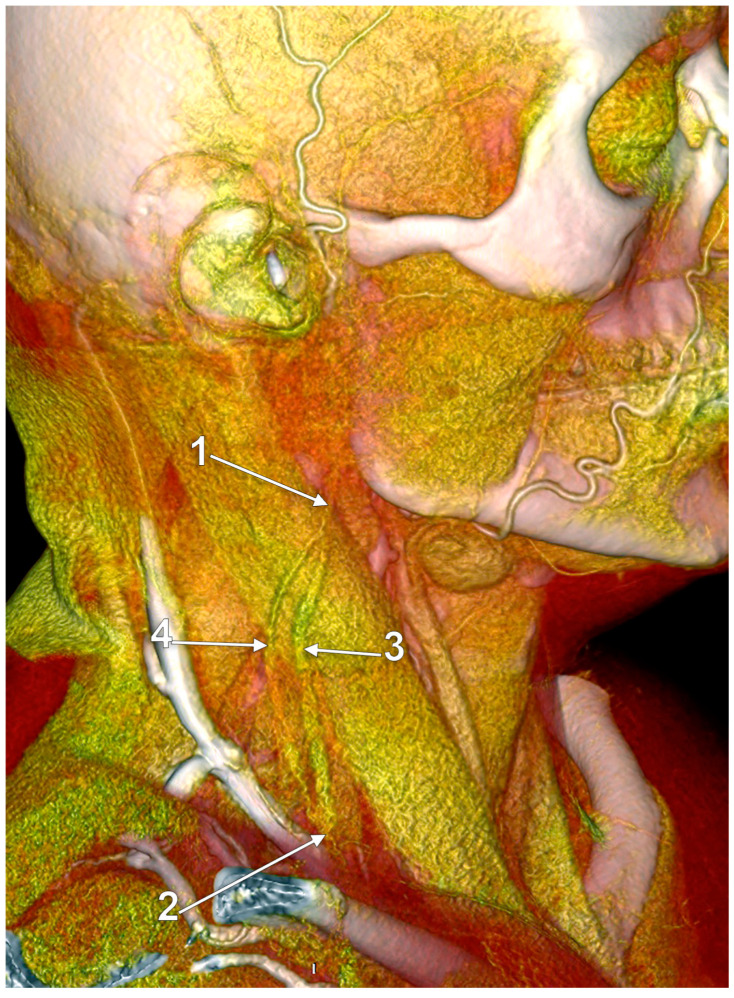
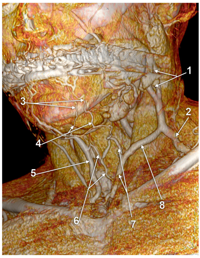
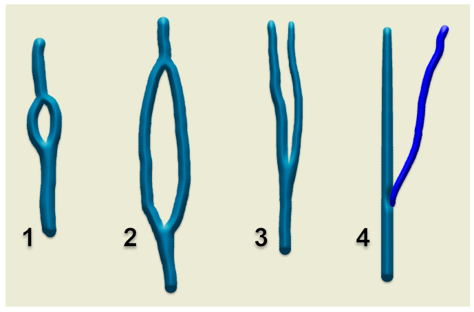
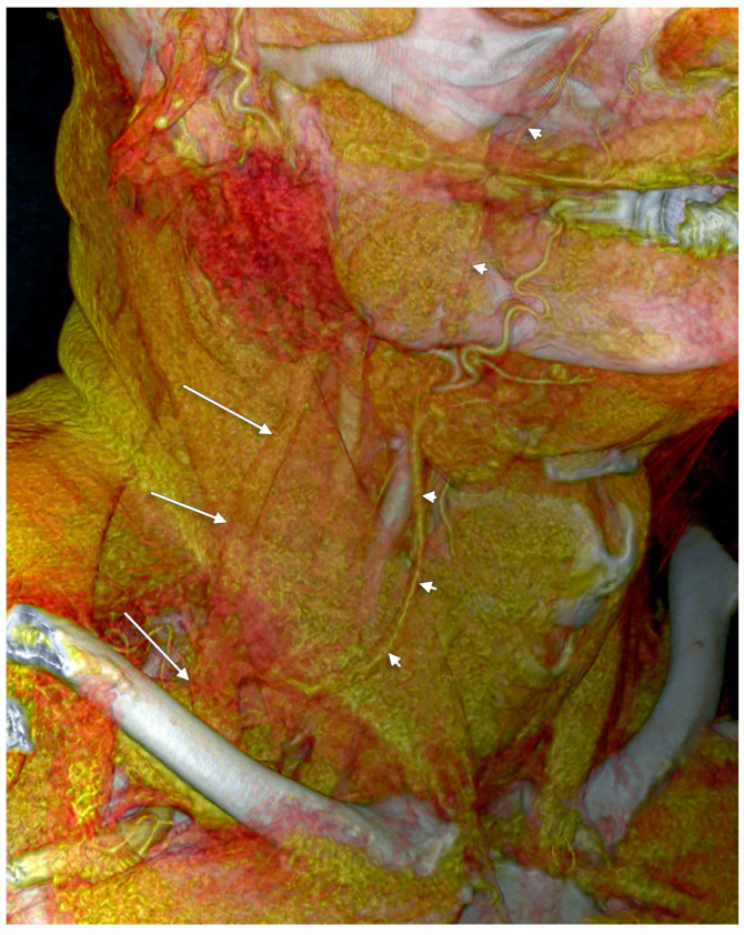
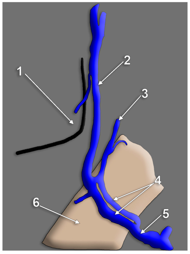
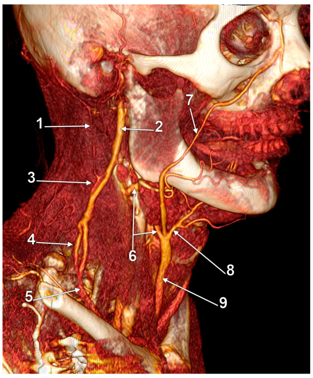
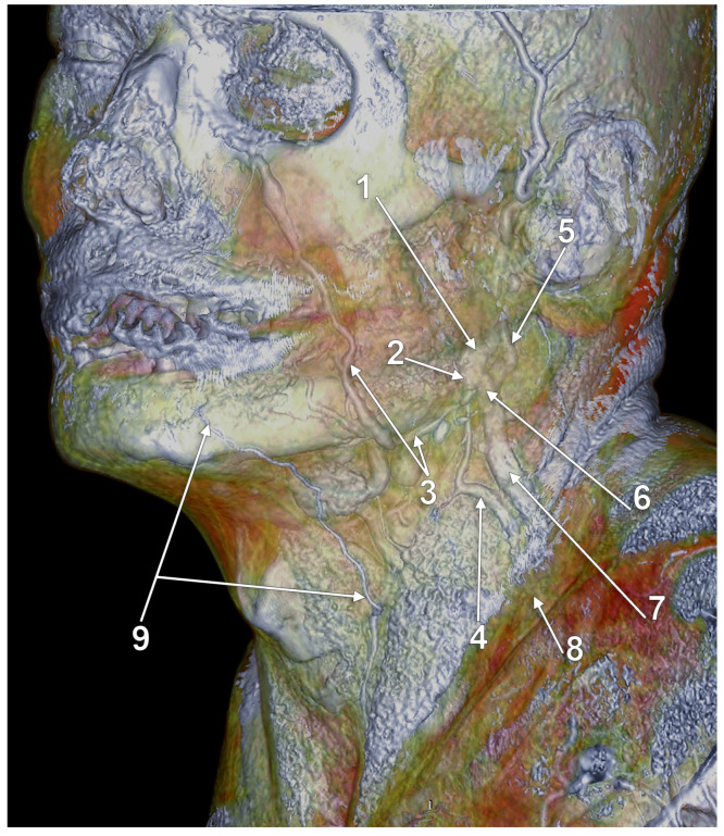
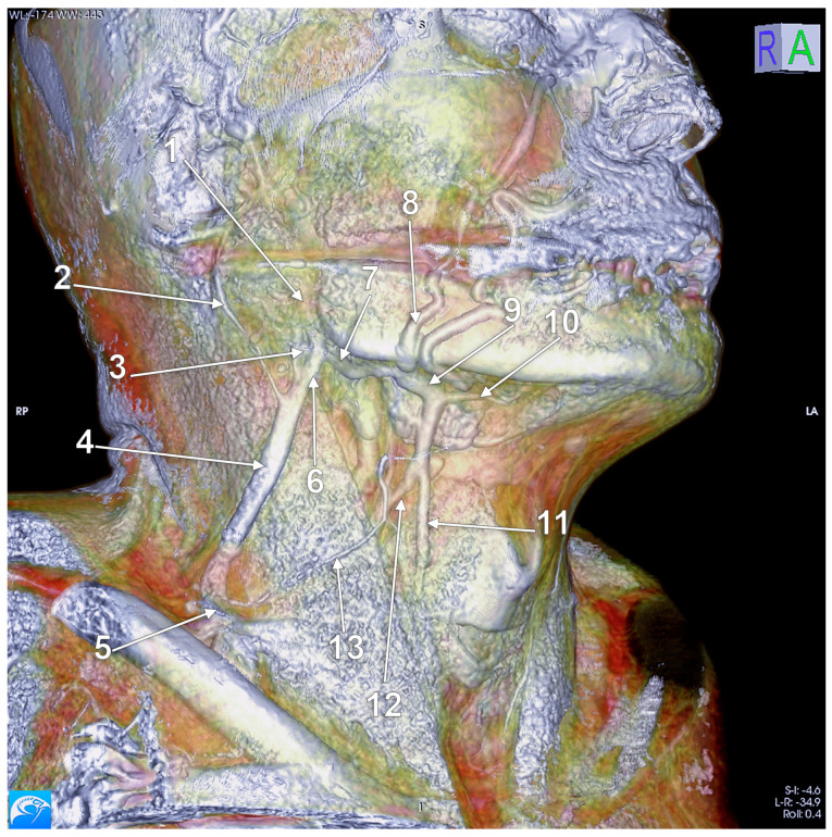
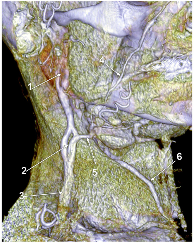
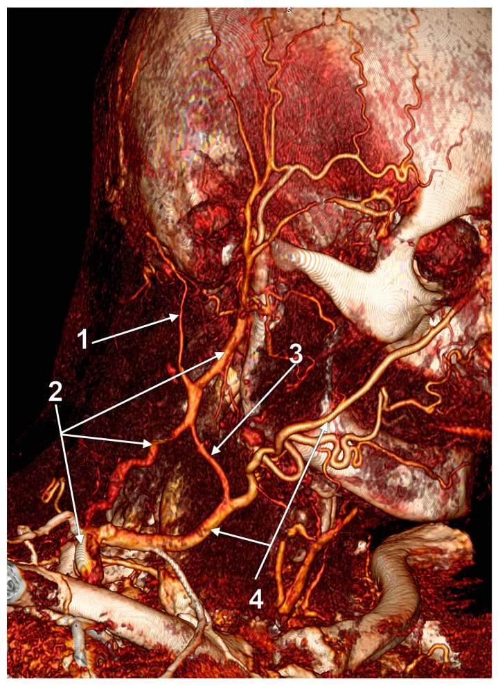
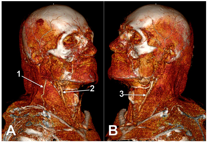
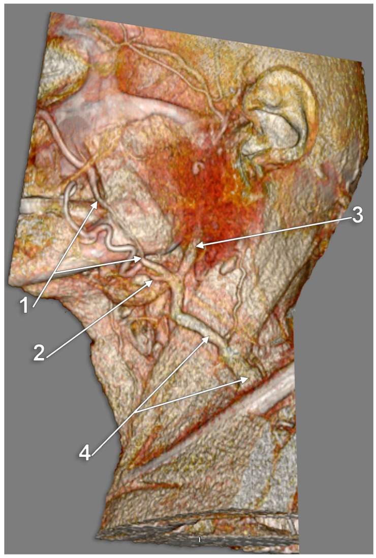
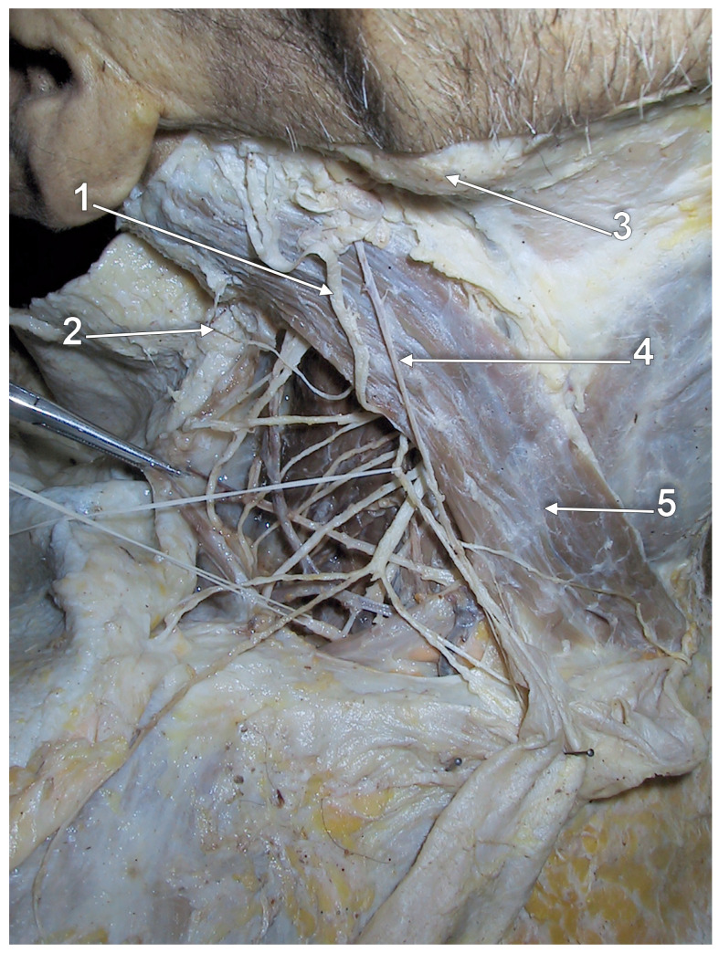
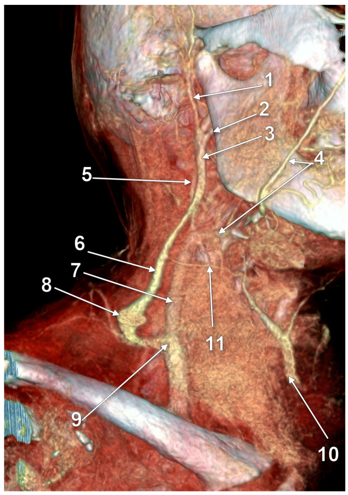
(1) Background: The external jugular vein (EJV) descends on the sternocleidomastoid muscle to drain deep into the subclavian vein. Anatomical variations of the EJV are relevant for identification of the greater auricular nerve, flap design and preparation, or EJV cannulation. (2) Methods: Different publications were comprehensively reviewed. Dissections and three-dimensional volume renderings of peculiar cases were used to sample the review. (3) Results: Different anatomical possibilities of the EJV were critically reviewed and documented: fenestrations and double fenestrations, true or false duplications, triplication, absence, aberrant origin or course, or bifurcation. Tributaries of the EJV, such as the facial and posterior external jugular veins, are discussed. The internal jugular vein termination of the EJV is also presented. (4) Conclusions: Care should be taken when different morphological features of the EJV are encountered or reported.

Keywords: computed tomography; duplication; fenestration; jugular veins; neck veins.


Conflict of interest statement

The authors declare no conflict of interest.

Figures

Figure 1
Figure 1
Right external jugular vein (EJV). Right side dissection, lateral view. 1. parotid gland; 2. lesser occipital nerve; 3. greater auricular nerve; 4. punctum nervosum; 5. transverse cervical nerve; 6. supraclavicular nerves; 7. sternocleidomastoid muscle; 8. communicating vein; 9. superficial cervical ansa; 10. cervical branch of the facial nerve; 11. EJV.
Figure 2
Figure 2
Right external jugular vein (EJV). Three-dimensional volume rendering (3D-VR). Right lateral view. 1. occipital vein; 2. EJV; 3. internal jugular vein; 4. sternocleidomastoid m.
Figure 3
Figure 3
Posterior external jugular vein coursing along the posterior border of the sternocleidomastoid muscle (3D-VR). Right lateral view. 1. parotid gland; 2. sternocleidomastoid m.; 3. external jugular vein; 4. posterior external jugular vein.
Figure 4
Figure 4
Posterior external jugular vein (PEJV) distanced from the sternocleidomastoid muscle (3D-VR). Left lateral view. 1. retromandibular vein; 2. anterior branch of the retromandibular vein; 3. external jugular vein; 4. PEJV.
Figure 5
Figure 5
Right external jugular vein (EJV) anastomosis with the left anterior jugular vein (AJV). Absent right AJV; 3D-VR, anterior view. 1. right facial vein; 2. right sternocleidomastoid muscle; 3. right EJV; 4. hyoid bone; 5. transverse hyoid anastomosis of the right EJV and left AJV; 6. left facial vein; 7. left sternocleidomastoid muscle; 8. left EJV; 9. left AJV deriving from the facial vein.
Figure 6
Figure 6
Large fenestration of the external jugular vein (EJV); 3D-VR, right lateral view. 1. undivided retromandibular vein; 2. sternocleidomastoid muscle; 3. facial vein; 4. posterior arm of the fenestrated EJV; 5. anterior arm of the fenestrated EJV; 6. common distal trunk of the EJV.
Figure 7
Figure 7
True duplication of the external jugular vein and fenestration of its posterior arm (3D-VR). Right lateral view. 1. parotid gland; 2. posterior auricular vein; 3. posterior arm of the duplicated external jugular vein (PEJV); 4. posterior external jugular vein; 5. fenestration of the PEJV; 6. common inferior trunk of the external jugular vein; 7. anterior arm of the duplicated external jugular vein (AEJV); 8. anterior jugular vein; 9. AEJV-to-anterior jugular communicating vein.
Figure 8
Figure 8
Anastomoses of the right external jugular and anterior jugular systems are depicted differently in Rouvière’s (A) and Pernkopf’s (B) drawings. Modified after [25,40]. 1. internal jugular vein; 2. subclavian vein; 3. brachiocephalic vein; 4. EJV; 5. AJV; 6. submental vein; 7. transverse anastomosis of the external jugular and anterior jugular veins; 8. oblique anastomosis of the retromandibular and anterior jugular veins.
Figure 9
Figure 9
The suprahyoid anastomosis of facial veins ((A), modified from [38]), compared with (B), the transverse hyoid anastomosis of the right external and left anterior jugular systems in Figure 5). 1. internal jugular vein; 2. subclavian vein; 3. external jugular vein; 4. facial vein; 5. anterior jugular vein; 6. submental/suprahyoid anastomosis of the facial veins; 7. transverse hyoid anastomosis of the right external and left anterior jugular systems (anatomic variant).
Figure 10
Figure 10
Large fenestration of the external jugular vein (EJV); 3D-VR, right lateral view. 1. proximal unique segment of the EJV; 2. distal unique segment of the EJV; 3. anterior arm of the EJV fenestration; 4. posterior arm of the EJV fenestration.
Figure 11
Figure 11
Fenestrated external jugular vein (3D-VR). Submental venous network draining the inferior labial veins. Anterior jugular network. Left antero-infero-lateral view. 1. proximal fenestration of the external jugular vein; 2. distal segment of the external jugular vein; 3. left inferior labial veins; 4. submental venous network; 5. right anterior jugular vein; 6. median cervical network; 7. left anterior jugular vein; 8. communicating vein.
Figure 12
Figure 12
Diagrams of morphologic possibilities of EJV fenestration or duplication. 1. small, slit-like fenestration; 2. large fenestration; 3. true duplication; 4. false duplication.
Figure 13
Figure 13
True duplication of the right external jugular vein. Trifurcated retromandibular vein. Prejugular anastomosis of the retromandibular and internal jugular veins (3D-VR). (A) Right lateral view. (B) Anterior view. 1. trifurcated retromandibular vein; 2. posterior arm of the duplicated external jugular vein; 3. anterior arm of the duplicated external jugular vein; 4. inferior common trunk of the external jugular vein; 5. facial vein; 6. anterior branch of the retromandibular vein (ARMV); 7. internal jugular vein (IJV); 8. superior thyroid vein; 9. prejugular ARMV-to-IJV anastomosis; 10. middle thyroid vein.
Figure 14
Figure 14
False duplication of the external jugular vein (3D-VR). Right antero-lateral view. The external jugular vein (arrows) descends on the sternocleidomastoid muscle. The facial vein (arrowheads) courses anteriorly and drains into the external jugular vein in the supraclavicular triangle.
Figure 15
Figure 15
Diagram of a left side venous variant reported as “duplication of the external jugular vein”, drawn after. In the original dissection picture, only the sternocleidomastoid muscle and nerves were labeled. 1. angle of mandible; 2. undivided retromandibular vein; 3. posterior auricular vein; 4. upper fenestrated segment of the external jugular vein; 5. supraclavicular segment of the external jugular vein; 6. sternocleidomastoid muscle.
Figure 16
Figure 16
Double fenestration of the external jugular vein (EJV). Trifurcated origin of the anterior jugular vein; 3D-VR, right lateral view. 1. occipital vein; 2. undivided retromandibular vein; 3. proximal EJV fenestration; 4. distal EJV fenestration; 5. terminal end of EJV; 6. internal jugular-to-anterior jugular communicating vein; 7. facial vein proper; 8. submental vein; 9. anterior jugular vein.
Figure 17
Figure 17
Duplicated retromandibular vein (RMV) continued as fenestrated external jugular vein (EJV); 3D-VR, left infero-lateral view. 1. RMV; 2. anterior division of the RMV; 3. facial vein proper; 4. anterior arm of the EJV fenestration, continuing (2); 5. posterior auricular vein; 6. posterior division of the RMV; 7. posterior arm of the EJV fenestration resulting from (6); 8. terminal end of the EJV; 9. inferior labial vein continued as anterior jugular vein.
Figure 18
Figure 18
Fenestrated retromandibular vein (3D-VR). Right lateral view. 1. retromandibular vein; 2. long posterior auricular vein; 3. fenestrated retromandibular vein (RMV); 4. external jugular vein (EJV); 5. terminal end of the EJV; 6. posterior division of the RMV; 7. anterior division of the RMV; 8. facial vein proper; 9. common facial vein; 10. submental vein; 11. anterior jugular vein; 12. distal segment of the common facial vein draining into the internal jugular vein; 13. superficial anterior cervical vein draining over the sternocleidomastoid muscle to the EJV.
Figure 19
Figure 19
Fenestrated undivided retromandibular vein (RMV). Fenestrated external jugular vein (EJV); 3D-VR, right lateral view. 1. RMV fenestration; 2. EJV fenestration; 3. distal EJV; 4. facial vein proper; 5. internal jugular-to-EJV communicating vein appears as a substitute of the anterior division of the RMV; 6. anterior jugular vein.
Figure 20
Figure 20
Complex venous architecture superficial to sternocleidomastoid muscle; 3D-VR, right antero-lateral view. 1. occipital vein; 2. external jugular vein; 3. communicating vein; 4. facial vein.
Figure 21
Figure 21
Absent left external jugular vein (3D-VR). (A) Right lateral view; (B) left lateral view. 1. right external jugular vein; 2. anterior branch of the right retromandibular vein continued as anterior jugular vein at the anterior border of the sternocleidomastoid muscle; 3. anterior branch of the left retromandibular vein continued as anterior jugular vein.
Figure 22
Figure 22
Facial vein continued as external jugular vein (3D-VR). Left side, infero-lateral view. 1. facial vein; 2. submental vein; 3. undivided retromandibular vein; 4. external jugular vein (variant).
Figure 23
Figure 23
Hypoplastic external jugular vein. Cadaver dissection. Right side, lateral view. 1. great auricular nerve; 2. lesser occipital nerve; 3. angle of mandible; 4. hypoplastic external jugular vein; 5. sternocleidomastoid muscle.
Figure 24
Figure 24
Internal jugular vein (IJV) drainage of an aneurysmal external jugular vein (EJV) via the Erb’s point (3D-VR). Right lateral view. 1. superficial temporal vein; 2. maxillary vein; 3. undivided retromandibular vein; 4. facial vein; 5. hypoplastic superior cervical segment of the IJV; 6. EJV; 7. normoplastic inferior cervical segment of the IJV; 8. EJV aneurysm; 9. EJV-to-IJV drainage; 10. anterior jugular vein (AJV); 11. EJV-AJV communicating vein.

References

    1. Gray H., Standring S., Anand N., Birch R., Collins P., Crossman A., Gleeson M., Jawaheer G., Smith A.L., Spratt J.D., et al. Gray’s Anatomy: The Anatomical Basis of Clinical Practice. Elsevier; London, UK: 2016.
    1. Lengele B., Hamoir M., Scalliet P., Gregoire V. Anatomical bases for the radiological delineation of lymph node areas. Major collecting trunks, head and neck. Radiother. Oncol. 2007;85:146–155. doi: 10.1016/j.radonc.2007.02.009. - DOI - PubMed
    1. Rusu M.C., Radoi P.M., Toader C. The transcerebral laterocavernous vein, a form of persisting primitive tentorial sinus. Surg. Radiol. Anat. 2022;44:1471–1474. doi: 10.1007/s00276-022-03038-y. - DOI - PubMed
    1. Minca D.I., Rusu M.C., Radoi P.M., Hostiuc S., Toader C. Transcallosal and Pericallosal Courses of the Anterior Cerebral Artery. Medicina. 2022;58:1365. doi: 10.3390/medicina58101365. - DOI - PMC - PubMed
    1. Minca D.I., Rusu M.C., Radoi P.M., Hostiuc S., Toader C. A New Classification of the Anatomical Variations of Labbe’s Inferior Anastomotic Vein. Tomography. 2022;8:2182–2192. doi: 10.3390/tomography8050183. - DOI - PMC - PubMed

LinkOut - more resources