. 2023 Feb 28;13(3):365.
doi: 10.3390/metabo13030365.

Data Independent Acquisition Reveals In-Depth Serum Proteome Changes in Canine Leishmaniosis

Affiliations

Data Independent Acquisition Reveals In-Depth Serum Proteome Changes in Canine Leishmaniosis

Franjo Martinković et al. Metabolites. .

Abstract

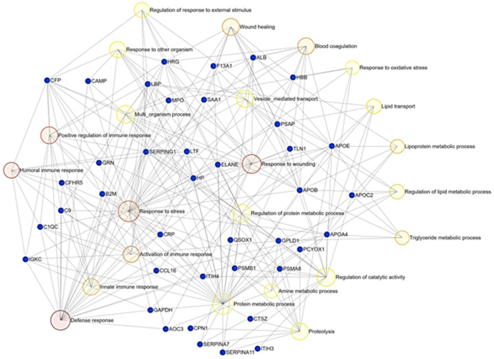
Comprehensive profiling of serum proteome provides valuable clues of health status and pathophysiological processes, making it the main strategy in biomarker discovery. However, the high dynamic range significantly decreases the number of detectable proteins, obstructing the insights into the underlying biological processes. To circumvent various serum enrichment methods, obtain high-quality proteome wide information using the next-generation proteomic, and study host response in canine leishmaniosis, we applied data-independent acquisition mass spectrometry (DIA-MS) for deep proteomic profiling of clinical samples. The non-depleted serum samples of healthy and naturally Leishmania-infected dogs were analyzed using the label-free 60-min gradient sequential window acquisition of all theoretical mass spectra (SWATH-MS) method. As a result, we identified 554 proteins, 140 of which differed significantly in abundance. Those were included in lipid metabolism, hematological abnormalities, immune response, and oxidative stress, providing valuable information about the complex molecular basis of the clinical and pathological landscape in canine leishmaniosis. Our results show that DIA-MS is a method of choice for understanding complex pathophysiological processes in serum and serum biomarker development.

Keywords: biomarker discovery; canine; data independent acquisition; leishmaniosis; mass spectrometry; proteomics; serum.


Conflict of interest statement

The authors declare no conflict of interest.

Figures

Figure 1
Figure 1
DDA-MS analysis reveals increase in number of identified serum protein groups (master proteins) after enrichment and fractionation (ProteoMiner and SCX fractions) compared to non-depleted sample without fractionation (Shotgun).
Figure 2
Figure 2
Spectral libraries generated by Spectronaut 16 Pulsar database search (in orange) and sample-specific library generated by Proteome Discoverer import (in blue). Both spectral libraries are provided through the Supplementary file S2.
Figure 3
Figure 3
(a) Principal component analysis (PCA) score plot of the second and first principal components (PC1 vs PC2) reveals the differences in serum proteomes from healthy (neg, in green) and Leishmania-infected dogs (pos, in red). (b) Volcano plot of DIA data depicted with the fold change of each differentially abundant protein and belonging q-value. Significantly differentially abundant proteins (q < 0.05) are highlighted in red (marked as Candidates).
Figure 4
Figure 4
Ridgeline diagram of enriched GO (database PANTHER: BP) showing the biological processes and log2FC of belonging deregulated proteins (red dots—up; blue dots—down) in serum of dogs with leishmaniosis. The shade of blue represents p-values as depicted. BP in grey is not significant. The GO diagram is created using ExpressAnalyst (https://www.expressanalyst.ca/ (accessed on 30 November 2022)) based on Welch’s t-test.
Figure 5
Figure 5
Bipartite network showing the results of enrichment analysis of deregulated genes (GO:MF, based on adjusted p-values ranked by Welch’s test) in serum of Leishmania-infected dogs compared to healthy ones performed using ExpressAnalyst (https://www.expressanalyst.ca/ (accessed on 30 November 2022)). Blue dots represent proteins, while yellow/red circles mark molecular functions affected by canine leishmaniosis.

Similar articles

Cited by

References

    1. Geyer P.E., Holdt L.M., Teupser D., Mann M. Revisiting biomarker discovery by plasma proteomics. Mol. Syst. Biol. 2017;13:942. doi: 10.15252/msb.20156297. - DOI - PMC - PubMed
    1. Fürsch J., Kammer K.-M., Kreft S.G., Beck M., Stengel F. Proteome-wide structural probing of low-abundant protein interactions by crosslinking mass spectrometry. Anal. Chem. 2020;92:4016–4022. doi: 10.1021/acs.analchem.9b05559. - DOI - PubMed
    1. Anderson N.L., Anderson N.G. The human plasma proteome: History, character, and diagnostic prospects. Mol. Cell. Proteom. 2002;1:845–867. doi: 10.1074/mcp.R200007-MCP200. - DOI - PubMed
    1. Paul J., Veenstra T.D. Separation of Serum and Plasma Proteins for In-Depth Proteomic Analysis. Separations. 2022;9:89. doi: 10.3390/separations9040089. - DOI
    1. Košiček M., Gudelj I., Horvatić A., Jović T., Vučković F., Lauc G., Hećimović S., Kosicek M., Gudelj I., Horvatic A., et al. N-glycome of the lysosomal glycocalyx is altered in Niemann-Pick Type C disease model cells. Mol. Cell. Proteom. 2018;17:631–642. doi: 10.1074/mcp.RA117.000129. - DOI - PMC - PubMed