. 2023 Mar 17;13(3):437.
doi: 10.3390/metabo13030437.

Overexpression of Bacterial Beta-Ketothiolase Improves Flax (Linum usitatissimum L.) Retting and Changes the Fibre Properties

Affiliations

Overexpression of Bacterial Beta-Ketothiolase Improves Flax (Linum usitatissimum L.) Retting and Changes the Fibre Properties

Justyna Mierziak et al. Metabolites. .

Abstract

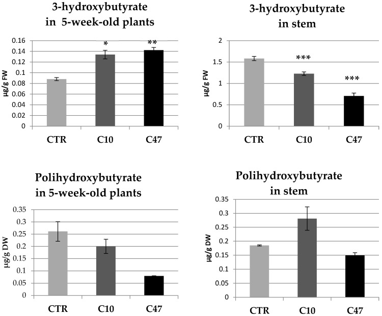
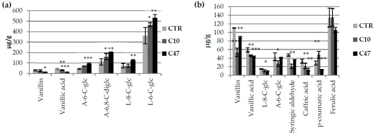
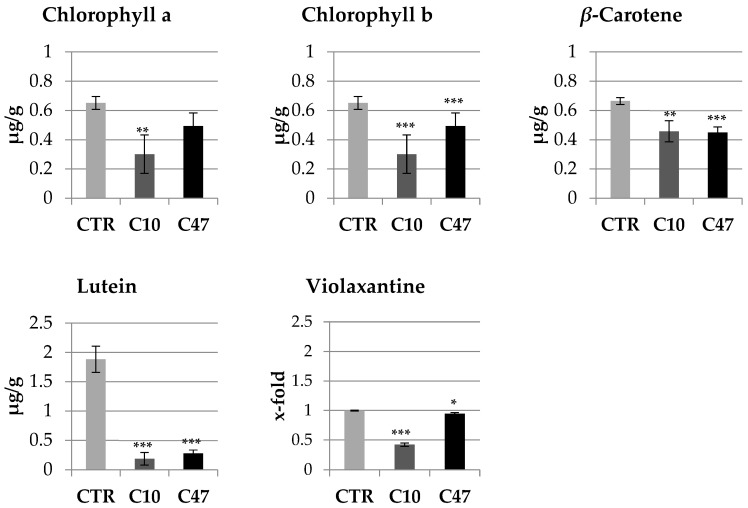
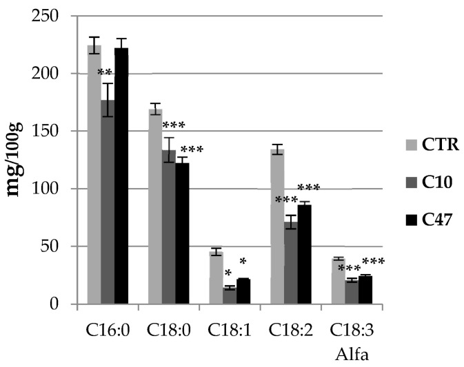
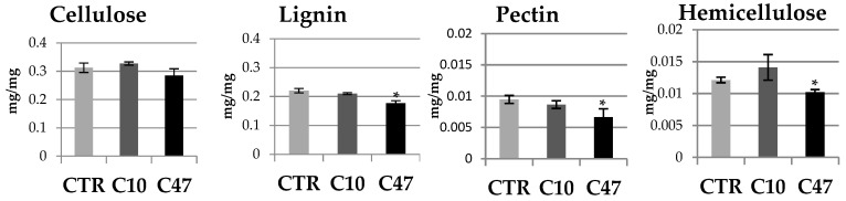
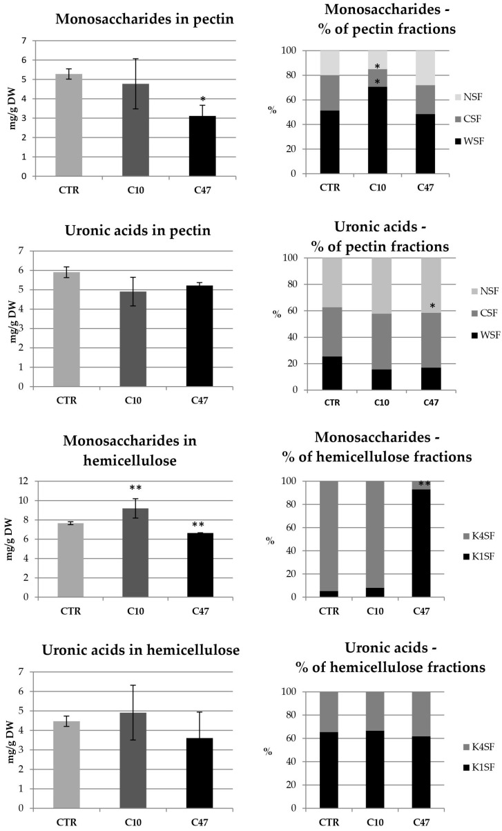
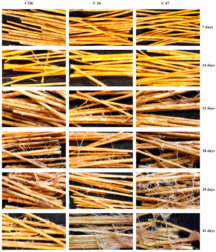
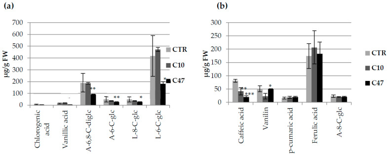
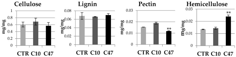
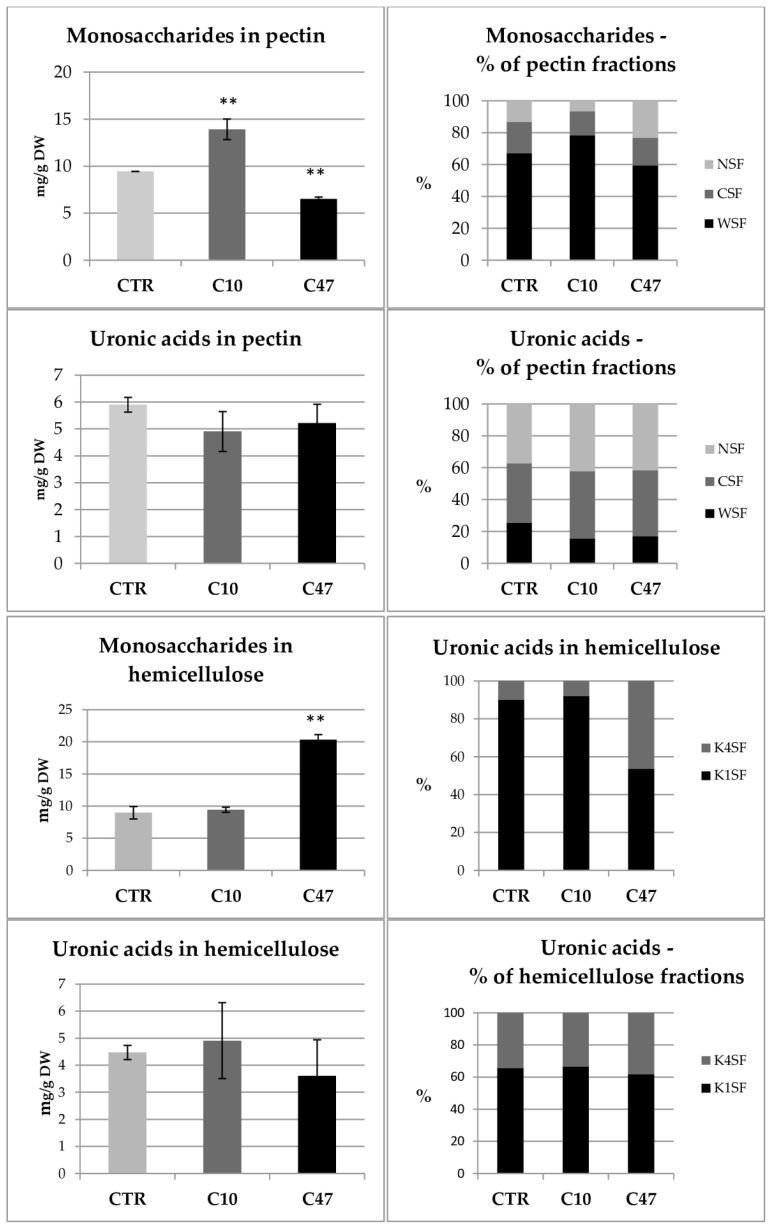
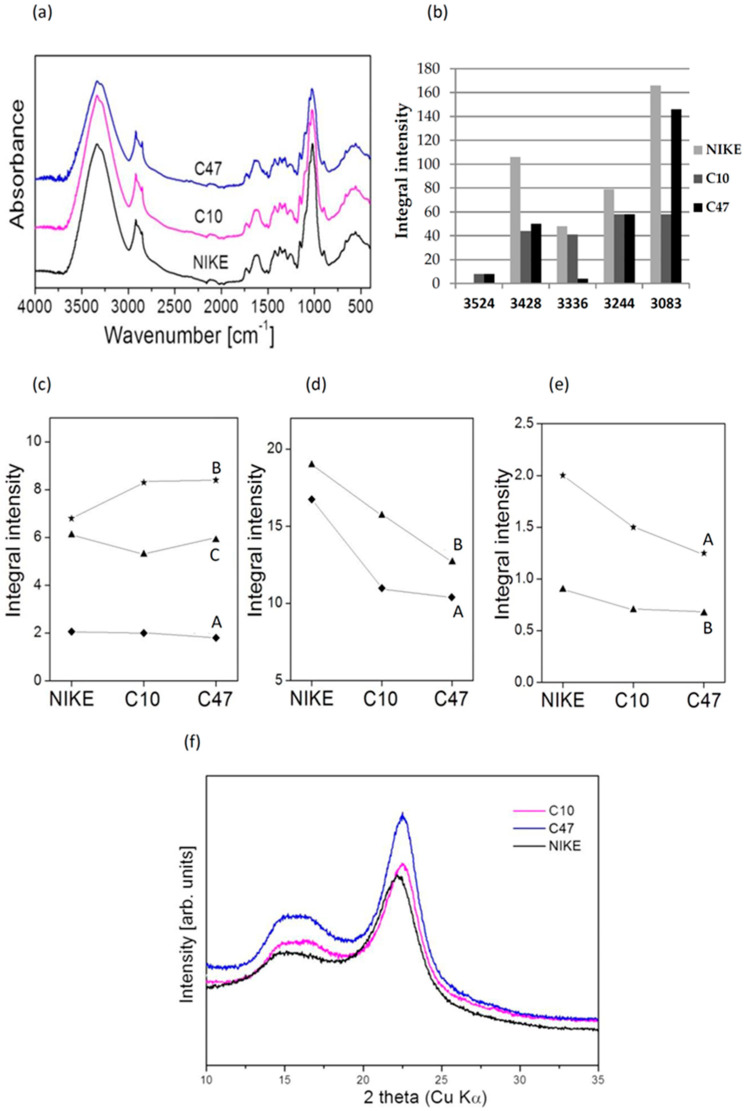
Beta-ketothiolases are involved in the beta-oxidation of fatty acids and the metabolism of hormones, benzenoids, and hydroxybutyrate. The expression of bacterial beta-ketothiolase in flax (Linum usitatissimum L.) results in an increase in endogenous beta-ketothiolase mRNA levels and beta-hydroxybutyrate content. In the present work, the effect of overexpression of beta-ketothiolase on retting and stem and fibre composition of flax plants is presented. The content of the components was evaluated by high-performance liquid chromatography, gas chromatography-mass spectrometry, Fourier-transform infrared spectroscopy, and biochemical methods. Changes in the stem cell walls, especially in the lower lignin and pectin content, resulted in more efficient retting. The overexpression of beta-ketothiolase reduced the fatty acid and carotenoid contents in flax and affected the distribution of phenolic compounds between free and cell wall-bound components. The obtained fibres were characterized by a slightly lower content of phenolic compounds and changes in the composition of the cell wall. Based on the IR analysis, we concluded that the production of hydroxybutyrate reduced the cellulose crystallinity and led to the formation of shorter but more flexible cellulose chains, while not changing the content of the cell wall components. We speculate that the changes in chemical composition of the stems and fibres are the result of the regulatory properties of hydroxybutyrate. This provides us with a novel way to influence metabolic composition in agriculturally important crops.

Keywords: 3-hydroxybutyrate; beta-ketothiolase; cell wall; flax fibre; flax stem; polyhydroxybutyrate.


Conflict of interest statement

The authors declare no conflict of interest.

Figures

Figure 1
Figure 1
3-hydroxybutyrate and polyhydroxybutyrate content in 5-week-old plants and stems in transgenic flax lines C10 and C47 and in non-transgenic flax (CTR). The results are depicted as the mean of three biological repetitions ± SD. Asterisks indicate statistically significant changes (*—p < 0.05, **—p < 0.01, ***—p < 0.001).
Figure 2
Figure 2
Levels of selected phenolic compounds in transgenic and control plants. Content of phenolic compounds in flax stem of maturation stage [(a)—free phenolic compounds: vanillin, vanillic acid, 6,8-C-diglucoside of apigenin (A-6,8-C-diglc), 6-C-glucose of luteolin (L-6-C-glc), 8-C-glucoside of luteolin (L-8-C-glc), and 6-C-glucoside of apigenin (A-6-C-glc) and (b)—bound phenolic compounds: vanillin, vanillic acid, syringaldehyde, luteolin 8-C-glucoside, apigenin 6-C-glucoside, p-coumaric acid, caffeic acid and ferulic acid] in transgenic flax lines C10 and C47, and control flax (CTR). The results are depicted as the mean of three biological repetitions ± SD. Asterisks indicate statistically significant changes (*—p < 0.05, **—p < 0.01, ***—p < 0.001).
Figure 3
Figure 3
Content of terpenoids in mature stems of transgenic flax lines C10 and C47 and control plants (CTR). The contents of chlorophyll a, chlorophyll b, beta-carotene, lutein, and violaxantine were obtained from the UPLC analysis. Calculations were performed using MassLynx 2.0 (Waters, Milford, CT, USA) software. The results are depicted as the mean of three biological repetitions ± SD. Asterisks indicate statistically significant changes (*—p < 0.05, **—p < 0.01, ***—p < 0.001).
Figure 4
Figure 4
Fatty acid contents in mature stem of transgenic flax lines C10 and C47 and control plants (CTR). Fatty acid (C16:0, C18:0, C18:1, C18:2, and C18:3 alfa) contents were determined using the GC-FID method. The results are depicted as the mean of three biological repetitions ± SD. Asterisks indicate statistically significant changes (*—p < 0.05, **—p < 0.01, ***—p < 0.001).
Figure 5
Figure 5
Contents of cellulose, lignin, pectin, and hemicellulose in the cell walls of mature stems of transgenic flax lines C10 and C47 and control (CTR). The results are depicted as the mean of three biological repetitions ± SD. Asterisks indicate statistically significant changes (*—p < 0.05).
Figure 6
Figure 6
Contents of monosaccharides and uronic acids in pectin and hemicellulose as well as percentage of monosaccharides and uronic acids in pectin and hemicellulose fractions in the cell walls (WSF, CSF, NSF, K1SF, and K4SF) of mature stems of transgenic flax lines C10 and C47 and control (CTR). The results are depicted as the mean of three biological repetitions ± SD. Asterisks indicate statistically significant changes (*—p < 0.05, **—p < 0.01).
Figure 7
Figure 7
Analysis of efficiency of retting for flax transgenic plants (C10 and C47) and control plants (CTR) after 7, 14, 21, 28, 35, and 41 days.
Figure 8
Figure 8
Levels of selected phenolic compounds in transgenic and control flax fibres. Content of phenolic compounds in fibres [(a)—free phenolic compounds: vanillic acid, chlorogenic acid, 6,8-C-diglucoside of apigenin (A-6,8-C-diglc), 6-C-glucose of luteolin (L-6-C-glc), 8-C-glucoside of luteolin (L-8-C-glc), and 6-C-glucoside of apigenin (A-6-C-glc) and (b)—bound phenolic compounds: vanillin, apigenin 8-C-glucoside (A-8-C-glc), p-coumaric acid, caffeic acid, and ferulic acid] from transgenic flax lines C10 and C47, and control flax (CTR). The results are depicted as the mean of three biological repetitions ± SD. Asterisks indicate statistically significant changes (*—p < 0.05, **—p < 0.01, ***—p < 0.001).
Figure 9
Figure 9
Content of cellulose, lignin, pectin, and hemicellulose in the cell wall of fibres of transgenic flax lines C10 and C47 and control (CTR). The results are depicted as the mean of three biological repetitions ± SD. Asterisks indicate statistically significant changes (**—p < 0.01).
Figure 10
Figure 10
Content of monosaccharides and uronic acids in pectin and hemicellulose as well as percentage of monosaccharides and uronic acids in pectin and hemicellulose fractions in the cell walls (WSF, CSF, NSF, K1SF, and K4SF) of fibres of transgenic flax lines C10 and C47 and control (CTR). The results are depicted as the mean of three biological repetitions ± SD. Asterisks indicate statistically significant changes (**—p < 0.01).
Figure 11
Figure 11
(a) IR spectra of fibres from transgenic flax lines C10 and C47 and non-transgenic flax Nike. (b) Wavenumber (ν) and integral intensity ratio of Lorentzian component related to the 2920 cm−1 standard (A) observed for non-transgenic (NIKE) and transgenic flax fibres. (c) Differences in the integral intensities of the bands at 1733 cm−1 (A), 1646 cm−1 (B), and 1600 cm−1 (C) for the NIKE, C10, and C47 samples. (d) Differences in the integral intensities of the bands at 1054 cm−1 (A) and 994 cm−1 (B) for the NIKE, C10, and C47 samples. (e) Differences in the integral intensities of the bands at 1507 cm−1 (A) and 1337 cm−1 (B) for the NIKE, C10, and C47 samples. (f) XRD diagrams of non-transgenic (NIKE) and transgenic flax samples.

Similar articles

References

    1. Preisner M., Wojtasik W., Kulma A., Żuk M., Szopa J. Kirk-Othmer Encyclopedia of Chemical Technology. John Wiley & Sons; Hoboken, NJ, USA: 2014. Flax Fiber.
    1. Dyminska L., Szatkowski M., Wrobel-Kwiatkowska M., Zuk M., Kurzawa A., Syska W., Gagor A., Zawadzki M., Ptak M., Maczka M., et al. Improved Properties of Micronized Genetically Modified Flax Fibers. J. Biotechnol. 2012;164:292–299. doi: 10.1016/j.jbiotec.2013.01.002. - DOI - PubMed
    1. Czemplik M., Mierziak J., Szopa J., Kulma A. Flavonoid C-Glucosides Derived from Flax Straw Extracts Reduce Human Breast Cancer Cell Growth In Vitro and Induce Apoptosis. Front. Pharmacol. 2016;7:282. doi: 10.3389/fphar.2016.00282. - DOI - PMC - PubMed
    1. Kulma A., Skórkowska-Telichowska K., Kostyn K., Szatkowski M., Skała J., Drulis-Kawa Z., Preisner M., Żuk M., Szperlik J., Wang Y.F., et al. New Flax Producing Bioplastic Fibers for Medical Purposes. Ind. Crops Prod. 2015;68:80–89. doi: 10.1016/j.indcrop.2014.09.013. - DOI
    1. Skórkowska-Telichowska K., Czemplik M., Kulma A., Szopa J. The Local Treatment and Available Dressings Designed for Chronic Wounds. J. Am. Acad. Dermatol. 2013;68:e117–e126. doi: 10.1016/j.jaad.2011.06.028. - DOI - PubMed

LinkOut - more resources