Core-Shell Particles: From Fabrication Methods to Diverse Manipulation Techniques
- PMID: 36984904
- PMCID: PMC10054063
- DOI: 10.3390/mi14030497
Core-Shell Particles: From Fabrication Methods to Diverse Manipulation Techniques
Abstract
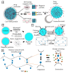
Core-shell particles are micro- or nanoparticles with solid, liquid, or gas cores encapsulated by protective solid shells. The unique composition of core and shell materials imparts smart properties on the particles. Core-shell particles are gaining increasing attention as tuneable and versatile carriers for pharmaceutical and biomedical applications including targeted drug delivery, controlled drug release, and biosensing. This review provides an overview of fabrication methods for core-shell particles followed by a brief discussion of their application and a detailed analysis of their manipulation including assembly, sorting, and triggered release. We compile current methodologies employed for manipulation of core-shell particles and demonstrate how existing methods of assembly and sorting micro/nanospheres can be adopted or modified for core-shell particles. Various triggered release approaches for diagnostics and drug delivery are also discussed in detail.
Keywords: assembly; digital microfluidics; sorting; targeted drug delivery; triggered release.
Conflict of interest statement
The authors have no conflicts to disclose.
Figures


Similar articles
-
Core-shell microparticles: From rational engineering to diverse applications.Adv Colloid Interface Sci. 2022 Jan;299:102568. doi: 10.1016/j.cis.2021.102568. Epub 2021 Nov 24. Adv Colloid Interface Sci. 2022. PMID: 34896747 Review.
-
Microfluidics for core-shell drug carrier particles - a review.RSC Adv. 2020 Dec 23;11(1):229-249. doi: 10.1039/d0ra08607j. eCollection 2020 Dec 21. RSC Adv. 2020. PMID: 35423057 Free PMC article. Review.
-
Facile fabrication of core-in-shell particles by the slow removal of the core and its use in the encapsulation of metal nanoparticles.Langmuir. 2008 May 6;24(9):4633-6. doi: 10.1021/la703955g. Epub 2008 Apr 15. Langmuir. 2008. PMID: 18410163
-
Core-Shell Magnetic Particles: Tailored Synthesis and Applications.Chem Rev. 2025 Jan 22;125(2):972-1048. doi: 10.1021/acs.chemrev.4c00710. Epub 2024 Dec 27. Chem Rev. 2025. PMID: 39729245 Review.
-
Compartmentalized and internally structured particles for drug delivery--a review.Curr Pharm Des. 2013;19(35):6298-314. doi: 10.2174/1381612811319350007. Curr Pharm Des. 2013. PMID: 23470000 Review.
Cited by
-
Microfluidic Gastrointestinal Cell Culture Technologies-Improvements in the Past Decade.Biosensors (Basel). 2024 Sep 19;14(9):449. doi: 10.3390/bios14090449. Biosensors (Basel). 2024. PMID: 39329824 Free PMC article. Review.
-
Enhanced Optical Management in Organic Solar Cells by Virtue of Square-Lattice Triple Core-Shell Nanostructures.Micromachines (Basel). 2023 Aug 9;14(8):1574. doi: 10.3390/mi14081574. Micromachines (Basel). 2023. PMID: 37630110 Free PMC article.
References
-
- Cao G., Wang Y. Nanostructures and Nanomaterials: Synthesis, Properties, and Applications. 2nd ed. World Scientific; Singapore: 2011.
-
- Astruc D. Nanoparticles and Catalysis. John Wiley & Sons; Hoboken, NJ, USA: 2008.
Publication types
Grants and funding
LinkOut - more resources
Full Text Sources
Other Literature Sources

