

Porous Structural Microfluidic Device for Biomedical Diagnosis: A Review
- PMID: 36984956
- PMCID: PMC10051279
- DOI: 10.3390/mi14030547
Porous Structural Microfluidic Device for Biomedical Diagnosis: A Review
Abstract
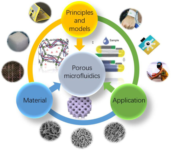
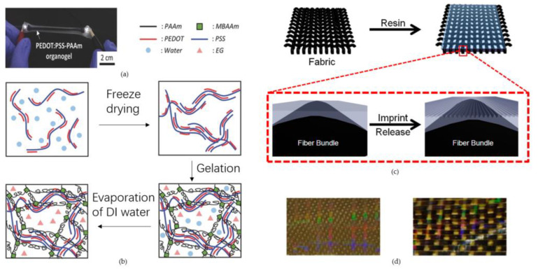
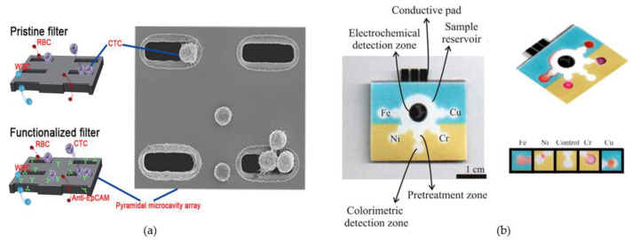
Microfluidics has recently received more and more attention in applications such as biomedical, chemical and medicine. With the development of microelectronics technology as well as material science in recent years, microfluidic devices have made great progress. Porous structures as a discontinuous medium in which the special flow phenomena of fluids lead to their potential and special applications in microfluidics offer a unique way to develop completely new microfluidic chips. In this article, we firstly introduce the fabrication methods for porous structures of different materials. Then, the physical effects of microfluid flow in porous media and their related physical models are discussed. Finally, the state-of-the-art porous microfluidic chips and their applications in biomedicine are summarized, and we present the current problems and future directions in this field.
Keywords: biomedical; biosensor; microfluidic; porous structure.
Conflict of interest statement
The authors declare no conflict of interest.
Figures
Similar articles
-
Microfluidic Platforms toward Rational Material Fabrication for Biomedical Applications.Small. 2020 Mar;16(9):e1903798. doi: 10.1002/smll.201903798. Epub 2019 Oct 25. Small. 2020. PMID: 31650698 Review.
-
Microfluidics devices for sports: A review on technology for biomedical application used in fields such as biomedicine, drug encapsulation, preparation of nanoparticles, cell targeting, analysis, diagnosis, and cell culture.Tissue Cell. 2024 Apr;87:102339. doi: 10.1016/j.tice.2024.102339. Epub 2024 Mar 2. Tissue Cell. 2024. PMID: 38432127 Review.
-
Recent advances in microfluidic-aided chitosan-based multifunctional materials for biomedical applications.Int J Pharm. 2021 May 1;600:120465. doi: 10.1016/j.ijpharm.2021.120465. Epub 2021 Mar 9. Int J Pharm. 2021. PMID: 33711469 Review.
-
Materials for microfluidic chip fabrication.Acc Chem Res. 2013 Nov 19;46(11):2396-406. doi: 10.1021/ar300314s. Epub 2013 Jun 11. Acc Chem Res. 2013. PMID: 24245999
-
[Applications of microfluidic paper-based chips in environmental analysis and detection].Se Pu. 2021 Aug;39(8):802-815. doi: 10.3724/SP.J.1123.2020.09004. Se Pu. 2021. PMID: 34212581 Free PMC article. Chinese.
Cited by
-
Development of a Tool for Verifying Leakage Detection in Microfluidic Systems.Micromachines (Basel). 2025 Jan 22;16(2):124. doi: 10.3390/mi16020124. Micromachines (Basel). 2025. PMID: 40047573 Free PMC article.
-
All-Printed Microfluidic-Electrochemical Devices for Glucose Detection.Biosensors (Basel). 2024 Nov 24;14(12):569. doi: 10.3390/bios14120569. Biosensors (Basel). 2024. PMID: 39727833 Free PMC article.
-
[Research progress on point-of-care testing of blood biochemical indexes based on microfluidic technology].Sheng Wu Yi Xue Gong Cheng Xue Za Zhi. 2025 Feb 25;42(1):205-211. doi: 10.7507/1001-5515.202406061. Sheng Wu Yi Xue Gong Cheng Xue Za Zhi. 2025. PMID: 40000194 Free PMC article. Review. Chinese.
References
-
- Zhang L., Tan Q., Fan J., Sun C., Luo Y., Liang R., Qiu J. Microfluidics for chiral separation of biomolecules. TrAC Trends Anal. Chem. 2023;158:116842. doi: 10.1016/j.trac.2022.116842. - DOI
-
- Terry S.C., Jerman J.H., Angell J.B. A gas chromatographic air analyzer fabricated on a silicon wafer. IEEE Trans. Electron Dev. 1979;26:1880–1886. doi: 10.1109/T-ED.1979.19791. - DOI
Publication types
Grants and funding
LinkOut - more resources
Full Text Sources
