. 2023 Feb 27;11(3):601.
doi: 10.3390/microorganisms11030601.

Effects of Cysticercus cellulosae Excretory-Secretory Antigens on the TGF-β Signaling Pathway and Th17 Cell Differentiation in Piglets, a Proteomic Analysis

Affiliations

Effects of Cysticercus cellulosae Excretory-Secretory Antigens on the TGF-β Signaling Pathway and Th17 Cell Differentiation in Piglets, a Proteomic Analysis

Wei He et al. Microorganisms. .

Abstract

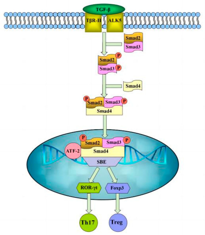
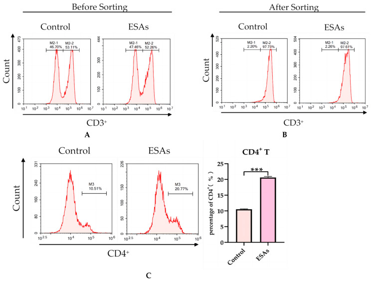
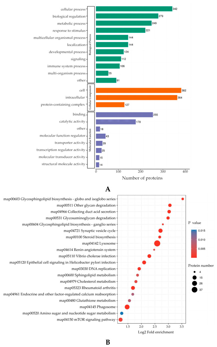
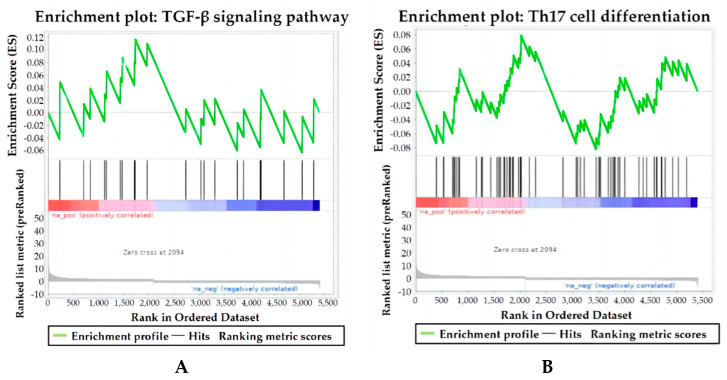
Excretory-secretory antigens (ESAs) of Cysticercus cellulosae can directly regulate the proliferation and differentiation of host T regulatory (Treg) cells, thus inhibiting host immune responses. However, previous studies have only focused on this phenomenon, and the molecular mechanisms behind the ways in which C. cellulosae ESAs regulate the differentiation of host Treg/Th17 cells have not been reported. We collected CD3+ T cells stimulated by C. cellulosae ESAs through magnetic bead sorting and used label-free quantification (LFQ) proteomics techniques to analyze the signaling pathways of C. cellulosae ESAs regulating Treg/Th17 cell differentiation. Through gene set enrichment analysis (GSEA), we found that C. cellulosae ESAs could upregulate the TGF-β signaling pathway and downregulate Th17 cell differentiation in piglet T cells. Interestingly, we also found that the IL-2/STAT5 signaling pathway also affects the downregulation of Th17 cell differentiation. C. cellulosae ESAs activate the TGF-β signaling pathway and the IL-2/STAT5 signaling pathway in host T cells to further regulate the differentiation of Treg/Th17 cells in order to evade host immune attack. This study lays the foundation for the subsequent verification of these pathways, and further clarifies the molecular mechanism of C. cellulosae-mediated immune evasion.

Keywords: TGF-β signaling pathway; Th17 cells differentiation; excretory–secretory antigens; proteomics.


Conflict of interest statement

The authors declare that the research was conducted in the absence of any commercial or financial relationships that could be construed as a potential conflict of interest.

Figures

Figure 1
Figure 1
TGF-β signaling pathway.
Figure 2
Figure 2
Analysis of the purity of CD3+ T cells and the ratio of CD4+ T cells. (A) The proportion of CD3+ T cells before magnetic bead sorting. (B) The proportion of CD3+ T cells after magnetic bead sorting. (C) Through flow cytometry analysis, the percentage of CD4+ T cells in the ESAs group was found to be significantly higher than that of the control group in terms of CD3+ T cells. *** p < 0.001.
Figure 3
Figure 3
Analysis of DEPs by GO functional annotation and KEGG signaling pathway enrichment. (A) GO enrichment elaborates the differential protein functions in terms of biological processes, cellular components, and molecular functions. (B) The KEGG pathway enrichment bubble plot showed the functional classification and pathway from significant enrichment (p value < 0.05) of DEPs.
Figure 4
Figure 4
Pathway enrichment map of TGF-β signaling pathway and Th17 cell differentiation by GSEA. The graph is divided into three parts, the first part is the line graph of the protein enrichment score (ES). In the top panel, the x-axis depicts all proteins, the y-axis depicts the running ES corresponding to the protein in the pathway, the peaks refer to the ES of the pathway, and the protein before the peak is the core protein of the pathway. For the middle panel, the pathway enrichment map refers to the hit, which marks the proteins located in this pathway with lines, and the color bars below indicate the corresponding expression amount of the proteins. The bottom panel shows the distribution plot of the quantitative value ratio for all proteins. (A) The enrichment map of the TGF-β signaling pathway is an upregulated pathway. (B) The enrichment map of the pathway for Th17 cells differentiation is a downregulated pathway.
Figure 5
Figure 5
PPI analysis was carried out for the enriched DEPs of the TGF- β signaling pathway and Th17 cell differentiation. Circles indicate DEPs, green circles are downregulated proteins and red circles are upregulated proteins.

References

    1. Himwaze C., Mucheleng’anga L.A., Telendiy V., Hamukale A., Tembo J., Kapata N., Ntoumi F., Zumla A. Cardiac cysticercosis and neurocysticercosis in sudden and unexpected community deaths in Lusaka, Zambia: A descriptive medico-legal post-mortem examination study. Int. J. Infect. Dis. 2022;115:195–200. doi: 10.1016/j.ijid.2021.11.042. - DOI - PubMed
    1. Pujari A., Bhaskaran K., Modaboyina S., Das D., Saluja G., Samdani A., Singh P., Bajaj M.S., Sharma N. Cysticercosis in ophthalmology. Surv. Ophthalmol. 2022;67:544–569. doi: 10.1016/j.survophthal.2021.07.002. - DOI - PubMed
    1. Prodjinotho U.F., Lema J., Lacorcia M., Schmidt V., Vejzagic N., Sikasunge C., Ngowi B., Winkler A.S., da Costa C.P. Host immune responses during Taenia solium Neurocysticercosis infection and treatment. PLoS Negl. Trop. Dis. 2020;14:e0008005. doi: 10.1371/journal.pntd.0008005. - DOI - PMC - PubMed
    1. Nyangi C., Stelzle D., Mkupasi E.M., Ngowi H.A., Churi A.J., Schmidt V., Mahonge C., Winkler A.S. Knowledge, attitudes and practices related to Taenia solium cysticercosis and taeniasis in Tanzania. BMC Infect. Dis. 2022;22:534. doi: 10.1186/s12879-022-07408-0. - DOI - PMC - PubMed
    1. Mendlovic F., Fleury A., Flisser A. Zoonotic Taenia infections with focus on cysticercosis due to Taenia solium in swine and humans. Res. Vet. Sci. 2021;134:69–77. doi: 10.1016/j.rvsc.2020.11.015. - DOI - PubMed

LinkOut - more resources