. 2023 Mar 3;11(3):657.
doi: 10.3390/microorganisms11030657.

Lactobacillus salivarius WZ1 Inhibits the Inflammatory Injury of Mouse Jejunum Caused by Enterotoxigenic Escherichia coli K88 by Regulating the TLR4/NF-κB/MyD88 Inflammatory Pathway and Gut Microbiota

Affiliations

Lactobacillus salivarius WZ1 Inhibits the Inflammatory Injury of Mouse Jejunum Caused by Enterotoxigenic Escherichia coli K88 by Regulating the TLR4/NF-κB/MyD88 Inflammatory Pathway and Gut Microbiota

Zhen Wei et al. Microorganisms. .

Abstract

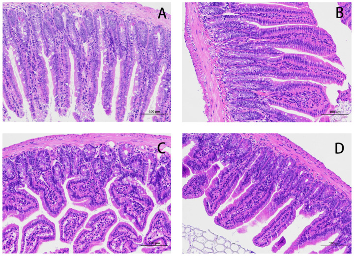
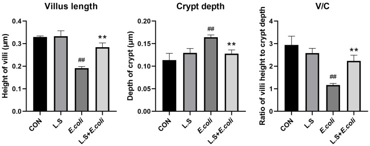
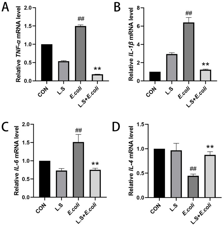
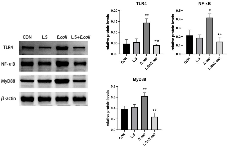
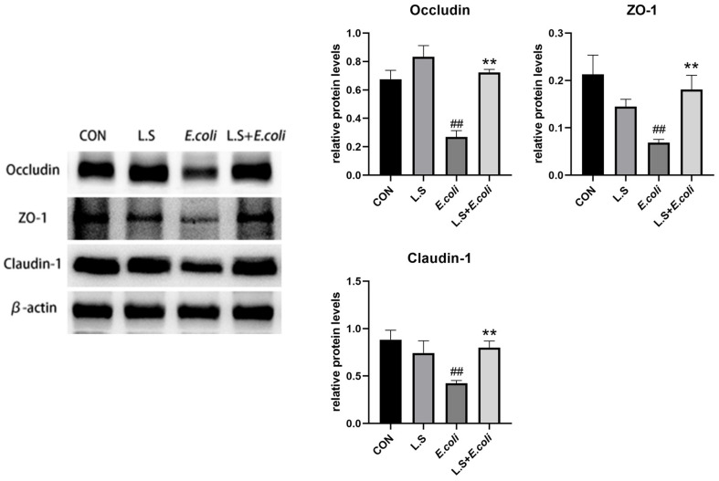
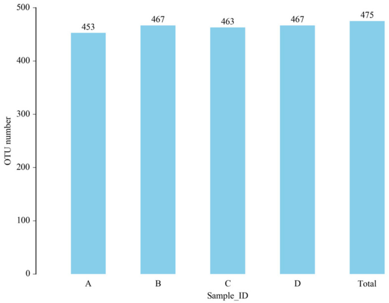
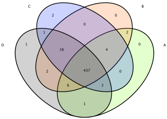
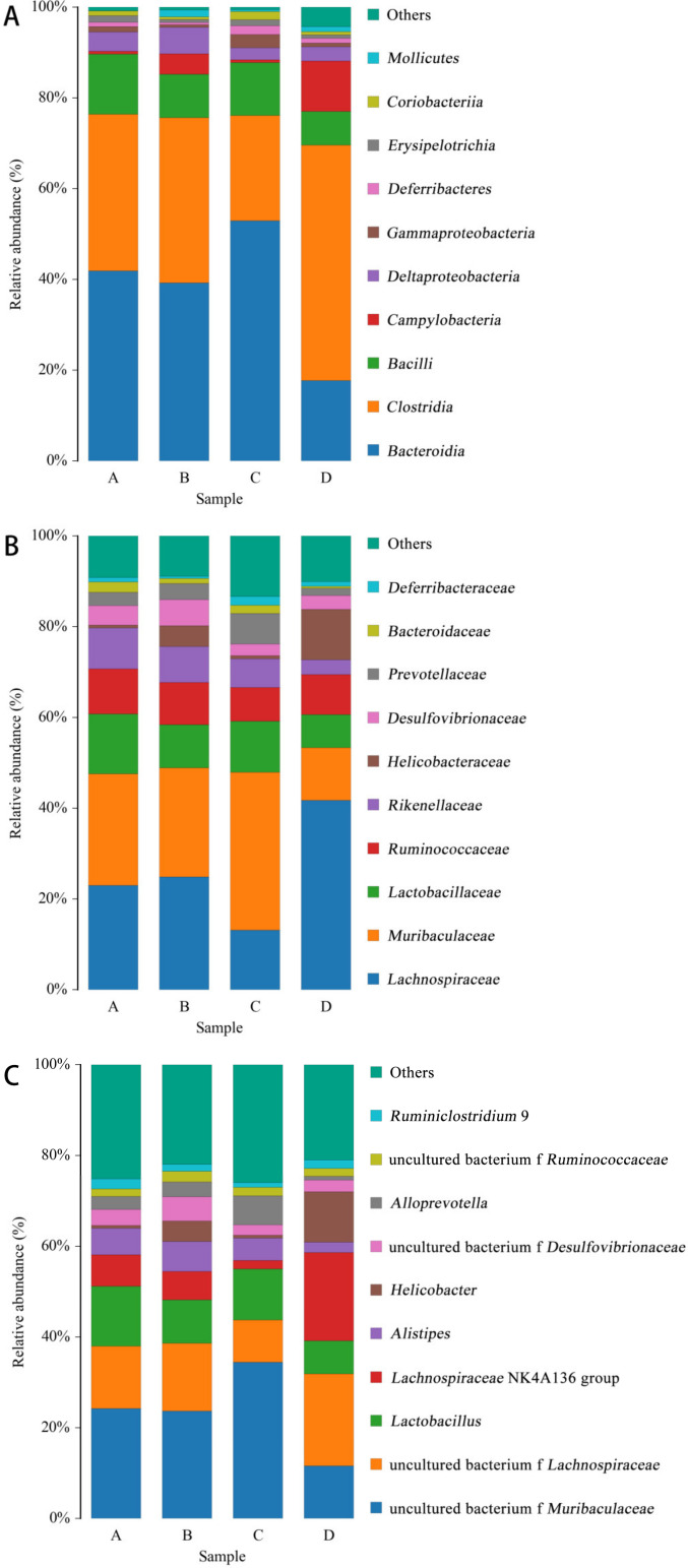
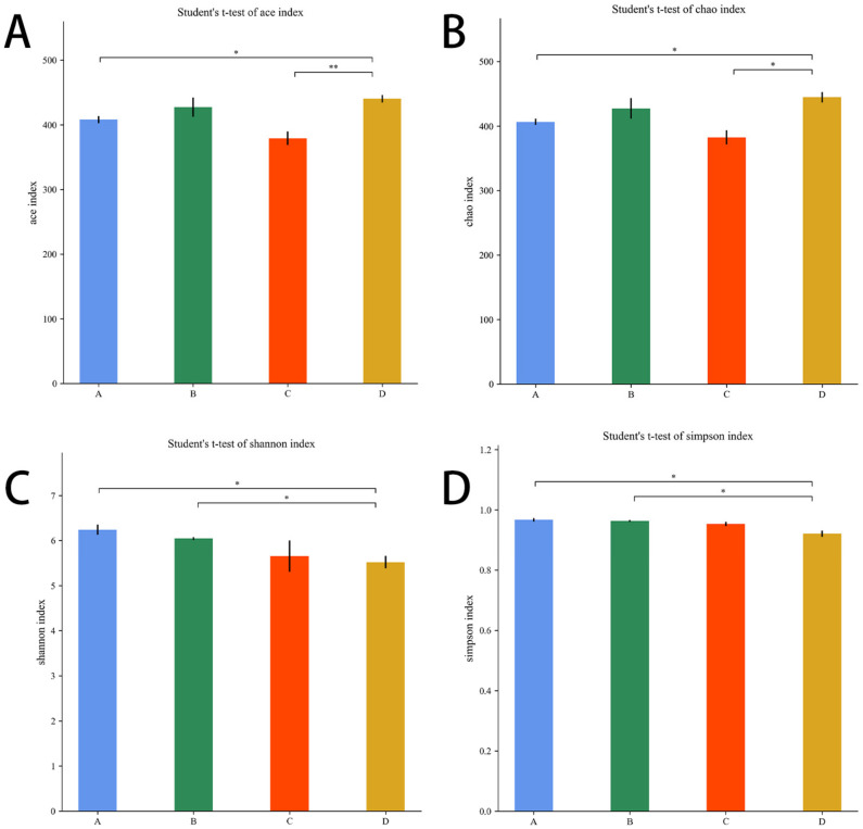
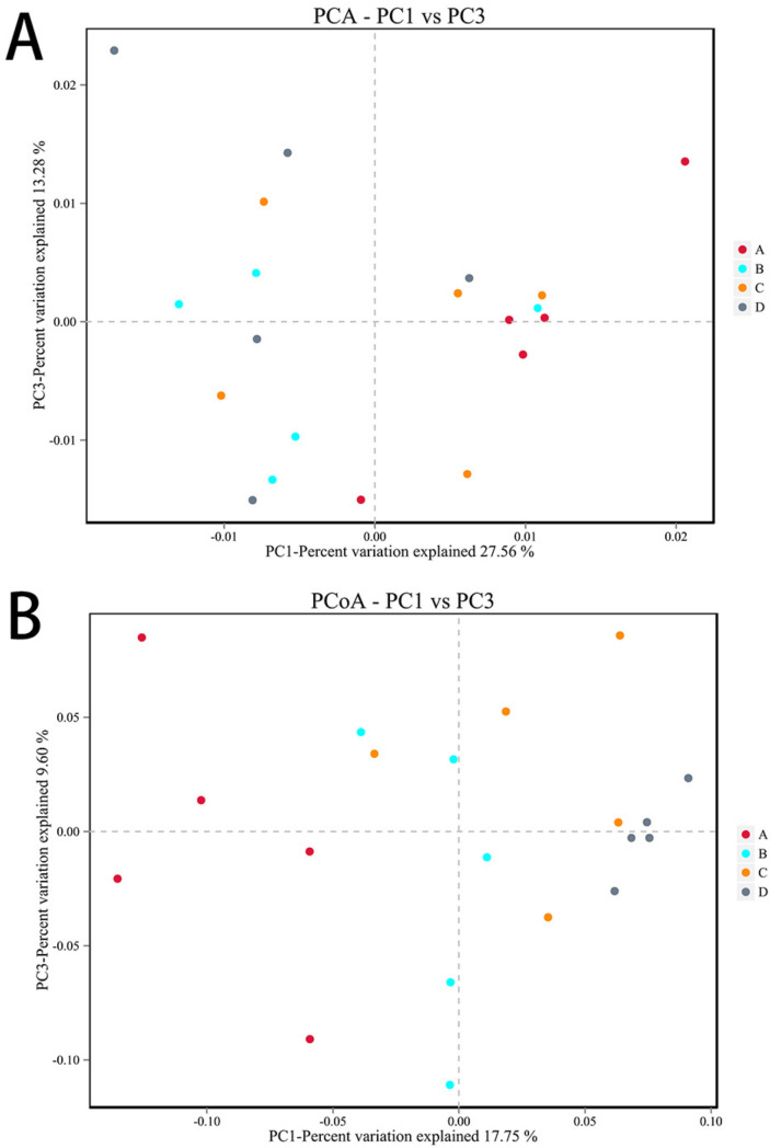
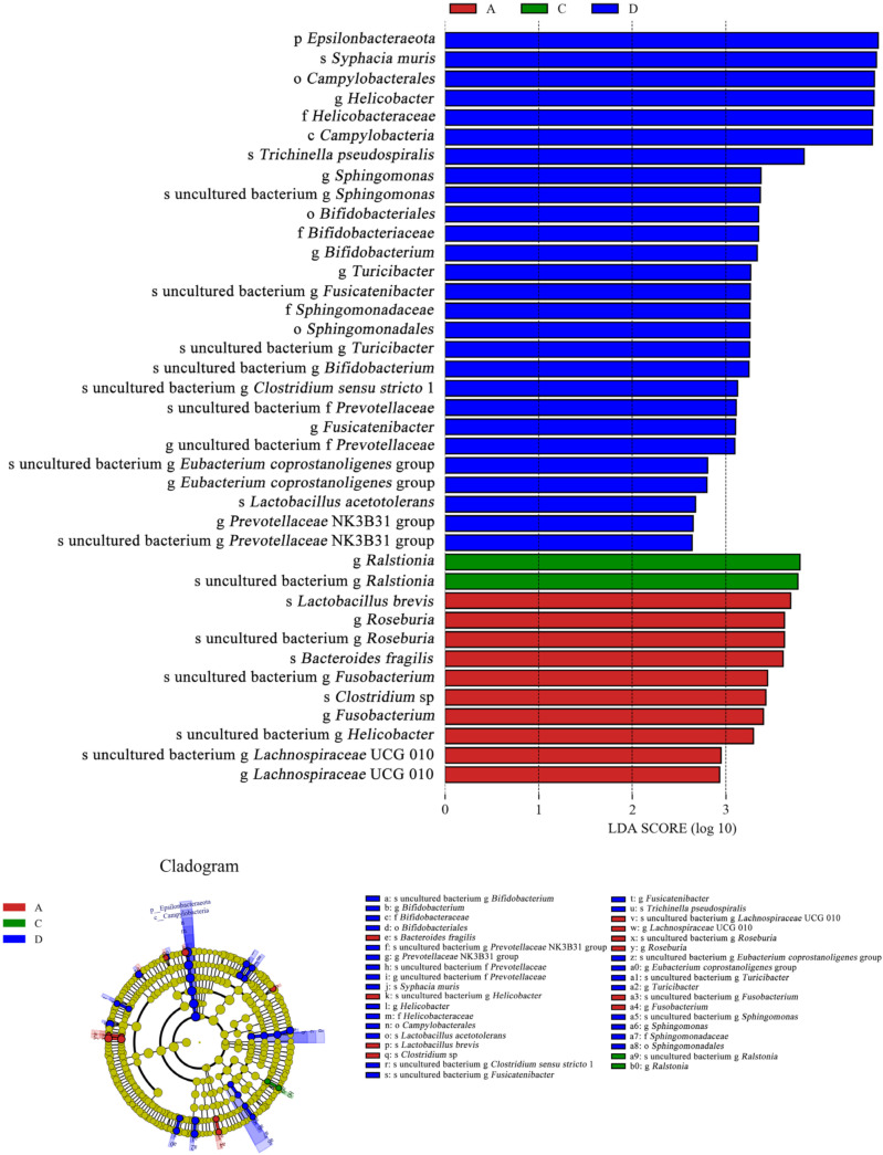
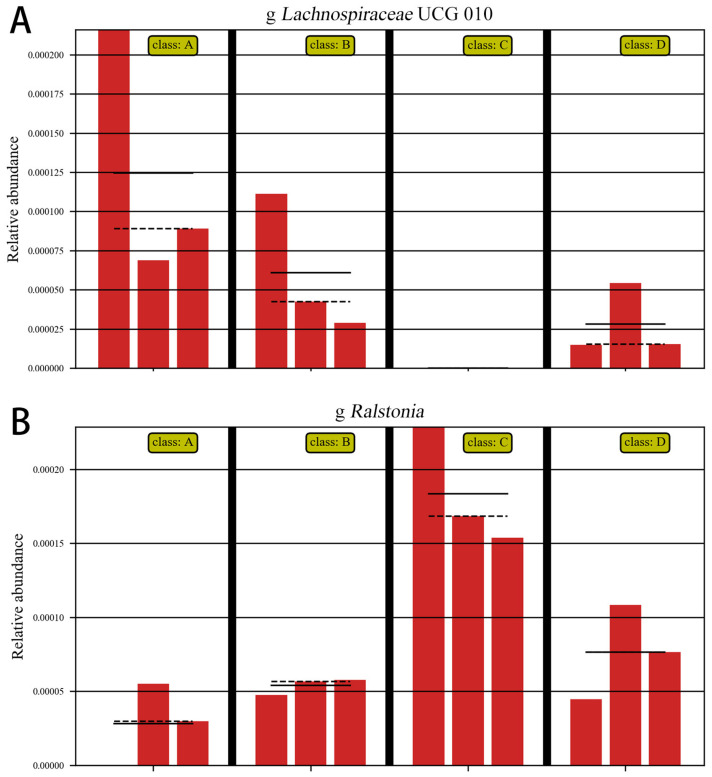
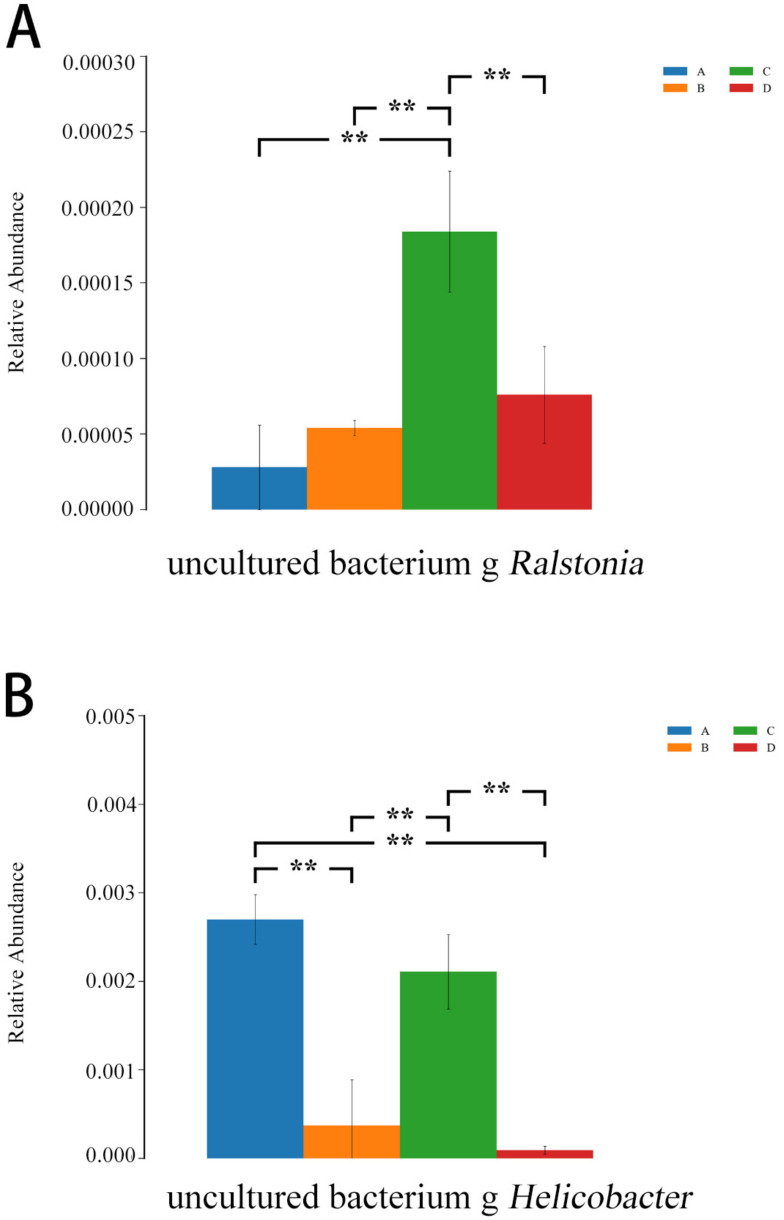
Replacing antibiotics with probiotics has become an important way to safely and effectively prevent and treat some gastrointestinal diseases. This study was conducted to investigate whether Lactobacillus salivarius WZ1 (L.S) could reduce the inflammatory injury to the mouse jejunum induced by Escherichia coli (ETEC) K88. Forty Kunming mice were randomly divided into four groups with 10 mice in each group. From day 1 to day 14, the control group and the E. coli group were administered with normal saline each day, while the L.S group and the L.S + E. coli group were gavaged with Lactobacillus salivarius WZ1 1 × 10⁸ CFU/mL each day. On the 15th day, the E. coli group and the L.S + E. coli group were intragastrically administered ETEC K88 1 × 10⁹ CFU/mL and sacrificed 24 h later. Our results show that pretreatment with Lactobacillus salivarius WZ1 can dramatically protect the jejunum morphological structure from the changes caused by ETEC K88 and relieve the morphological lesions of the jejunum, inhibiting changes in the mRNA expressions of TNF-α, IL-1β and IL-6 and the protein expressions of TLR4, NF-κB and MyD88 in the intestinal tissue of mice caused by ETEC K88. Moreover, pretreatment with Lactobacillus salivarius WZ1 also increased the relative abundance of beneficial genera such as Lactobacillus and Bifidobacterium and decreased the abundance of harmful genera such as Ralstonia and Helicobacter in the gut. These results demonstrate that Lactobacillus salivarius WZ1 can inhibit the inflammatory damage caused by ETEC K88 in mouse jejunum by regulating the TLR4/NF-κB/MyD88 inflammatory pathway and gut microbiota.

Keywords: ETEC K88; Lactobacillus salivarius; TLR4/NF-κB/MyD88; gut microbiota; inflammatory injury; mice.


Conflict of interest statement

The authors declare no conflict of interest.

Figures

Figure 1
Figure 1
HE staining of mouse jejunum tissue (200×). Note: (A): CON group, (B): L.S group, (C): E. coli group, (D): L.S + E. coli group.
Figure 2
Figure 2
Villus length and crypt depth in mouse jejunum tissue and the ratio between them. Note: ## in the figure indicates a very significant difference compared with the CON group (p < 0.01); ** indicates a very significant difference compared with the E. coli group (p < 0.01).
Figure 3
Figure 3
Relative expression of inflammatory factor mRNA in mouse jejunum tissue. Note: ## in the figure indicates a very significant difference compared with the CON group (p < 0.01); ** indicates a very significant difference compared with the E. coli group (p < 0.01). (A): Relative TNF-α mRNA expression, (B): Relative IL-1β mRNA expression, (C) Relative IL-6 mRNA expression, (D) Relative IL-4 mRNA expression.
Figure 4
Figure 4
Expression of inflammation-related proteins in mouse jejunum. Note: # in the figure indicates a significant difference compared with the CON group (p < 0.05); ## indicates a very significant difference compared with the CON group (p < 0.01); ** indicates a difference with the E. coli group where the difference is extremely significant (p < 0.01).
Figure 5
Figure 5
Expression of tight-junction-related proteins in the intestinal epithelium of the mouse jejunum. Note: ## in the figure indicates a very significant difference compared with the CON group (p < 0.01); ** indicates a very significant difference compared with the E. coli group (p < 0.01).
Figure 6
Figure 6
Number of OTUs. Note: A: CON group, B: L.S group, C: E. coli group, D: L.S + E. coli group.
Figure 7
Figure 7
Venn diagram. Note: A: CON group, B: L.S group, C: E. coli group, D: L.S + E. coli group.
Figure 8
Figure 8
Species distribution histogram. Note: A: CON group, B: L.S group, C: E.coli group, D: L.S + E. coli group. (A): class level. (B): family level. (C): genus level.
Figure 9
Figure 9
Alpha diversity index. Note: A: CON group, B: L.S group, C: E. coli group, D: L.S + E. coli group. * indicates significant difference (p < 0.05); ** indicates extremely significant difference (p < 0.01). (A): Ace index. (B): Chao1 index. (C): Shannon index .(D): Simpson index.
Figure 10
Figure 10
Beta diversity analysis. Note: Each data point represents a different sample (its color representing the group to which it belongs). The abscissa (ordinate) represents the first (second) principal component. The percentages shown are the contributions the component makes to the sample difference. A: CON group, B: L.S group, C: E. coli group, D: L.S + E. coli group. (A): PCA analyses (B): PCoA analyses.
Figure 11
Figure 11
LEfSe analysis. Note: A: CON group, C: E. coli group, D: L.S + E. coli group.
Figure 12
Figure 12
Abundance comparison chart. Note: A: CON group, B: L.S group, C: E. coli group, D: L.S + E. coli group. (A): the abundance of Lachnospiraceae (B): the abundance of Ralstonia.
Figure 13
Figure 13
ANOVA analysis diagram. Note: A: CON group, B: L.S group, C: E. coli group, D: L.S + E. coli group. ** indicates extremely significant difference (p < 0.01). (A): the relative abundances of Ralstonia (B): the relative abundances of Helicobacter.

Similar articles

Cited by

References

    1. Viidu D.A., Motus K. Implementation of a pre-calving vaccination programme against rotavirus, coronavirus and enterotoxigenic Escherichia coli (F5) and association with dairy calf survival. BMC Vet. Res. 2022;18:59. doi: 10.1186/s12917-022-03154-2. - DOI - PMC - PubMed
    1. Foster D.M., Smith G.W. Pathophysiology of diarrhea in calves. Vet. Clin. N. Am. Food Anim. Pract. 2009;25:13–36. doi: 10.1016/j.cvfa.2008.10.013. - DOI - PMC - PubMed
    1. Fesseha H., Mathewos M., Aliye S., Mekonnen E. Isolation and antibiogram of Escherichia coli O157: H7 from diarrhoeic calves in urban and peri-urban dairy farms of Hawassa town. Vet. Med. Sci. 2022;8:864–876. doi: 10.1002/vms3.686. - DOI - PMC - PubMed
    1. Cho Y.I., Yoon K.J. An overview of calf diarrhea—Infectious etiology, diagnosis, and intervention. J. Vet. Sci. 2014;15:1–17. doi: 10.4142/jvs.2014.15.1.1. - DOI - PMC - PubMed
    1. Prieto A., Lopez-Novo C., Diaz P., Diaz-Cao J.M., Lopez-Lorenzo G., Anton C., Remesar S., Garcia-Dios D., Lopez C., Panadero R., et al. Antimicrobial Susceptibility of Enterotoxigenic Escherichia coli from Diarrhoeic Neonatal Calves in Spain. Animals. 2022;12:264. doi: 10.3390/ani12030264. - DOI - PMC - PubMed

LinkOut - more resources