

The "Bald Disease" of the Sea Urchin Paracentrotus lividus: Pathogenicity, Molecular Identification of the Causative Agent and Therapeutic Approach
- PMID: 36985336
- PMCID: PMC10056887
- DOI: 10.3390/microorganisms11030763
The "Bald Disease" of the Sea Urchin Paracentrotus lividus: Pathogenicity, Molecular Identification of the Causative Agent and Therapeutic Approach
Abstract
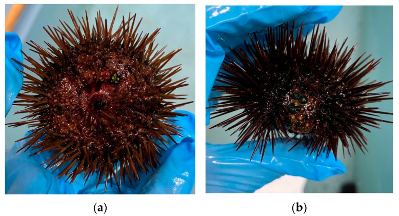
In recent decades, various species of Mediterranean sea urchins, including Paracentrotus lividus, have been subject to widespread seasonal episodes of mass mortality whose causative agents are still unclear. In particular, P. lividus is subject to late winter events of mortality, due to a disease manifested by a massive loss of spines and the presence of greenish amorphous material on the tests (i.e., the sea urchin skeleton consisting of spongeous calcite). Documented mortality events show a seasonal epidemic diffusion and might produce economic losses also in aquaculture facilities, besides the environmental constraints to its diffusion. We collected individuals showing conspicuous lesions on the body surface and reared them in recirculated aquaria. Samples of external mucous were collected along with coelomic liquids and cultured to isolate bacterial and fungal strains, further submitted to molecular identification through the amplification of prokaryotic 16S rDNA. In addition, pools of infected sea urchins were reared in recirculated tanks after short baths in a formulated therapeutic compound and their survival rates were compared to non-treated individuals for variable periods. Here, we aimed at a redescription of the etiopathogenetic nature of the parasites and tested the efficacy of a possible treatment, to be proposed for aquaculture purposes.
Keywords: bacteria; bald disease; echinoid necrosis; epidemics; sea urchin.
Conflict of interest statement
The authors declare no conflict of interest.
Figures


References
-
- Guerinot M.L., Patriquin D.G. The association of N2-fixing bacteria with sea urchins. Mar. Biol. 1981;62:197–207. doi: 10.1007/BF00388183. - DOI
-
- Holland N.D., Nealson K.H. The fine structure of the echinoderm cuticle and the subcuticular associated bacteria of echinoderms. Acta Zool. 1978;59:169–185. doi: 10.1111/j.1463-6395.1978.tb01032.x. - DOI
-
- Kaneshiro E.S., Karp R.D. The ultrastructure of coelomocytes of the sea star Dermasterias imbricata. Biol. Bull. 1980;159:295–310. doi: 10.2307/1541094. - DOI
-
- Wardlaw A.C., Unkles S.E. Bactericidal activity of coelomic fluid from the sea urchin Echinus esculentus. J. Invertebr. Pathol. 1978;32:25–34. doi: 10.1016/0022-2011(78)90170-2. - DOI
LinkOut - more resources
Full Text Sources
Miscellaneous
