. 2023 Mar 16;28(6):2691.
doi: 10.3390/molecules28062691.

Novel Steroidal[17,16- d]pyrimidines Derived from Epiandrosterone and Androsterone: Synthesis, Characterization and Configuration-Activity Relationships

Affiliations

Novel Steroidal[17,16- d]pyrimidines Derived from Epiandrosterone and Androsterone: Synthesis, Characterization and Configuration-Activity Relationships

Fei Yang et al. Molecules. .

Abstract

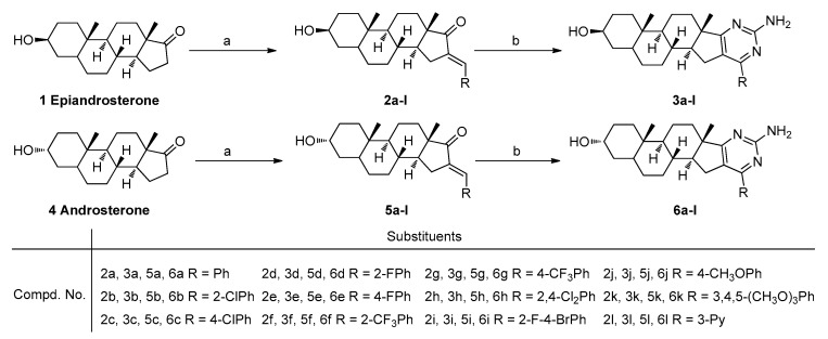
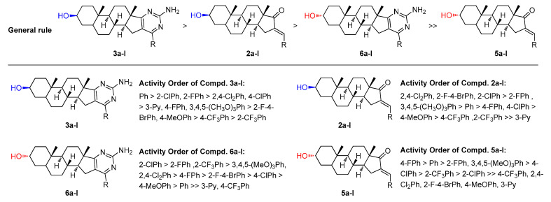
Two series of novel steroidal[17,16-d]pyrimidines derived from natural epiandrosterone and androsterone were designed and synthesized, and these compounds were screened for their potential anticancer activities. The preliminary bioassay indicated that some of these prepared compounds exhibited significantly good cytotoxic activities against human gastric cancer (SGC-7901), lung cancer (A549), and hepatocellular liver carcinoma (HepG2) cell lines compared with 5-fluorouracil (5-FU), epiandrosterone, and androsterone. Especially the respective pairs from epiandrosterone and androsterone showed significantly different inhibitory activities, and the possible configuration-activity relationships have also been summarized and discussed based on kinase assay and molecular docking, which indicated that the inhibition activities of these steroidal[17,16-d]pyrimidines might obviously be affected by the configuration of the hydroxyl group in the part of the steroidal scaffold.

Keywords: SARs; androsterone; bioactivity; epiandrosterone; molecular docking; steroidal[17,16-d]pyrimidines; synthesis.


Conflict of interest statement

The authors declare no conflict of interest.

Figures

Figure 1
Figure 1
Chemical structures of typical compounds with antiproliferative activity (A) epiandrosterone derivatives; (B) 2-aminopyrimidine derivatives; (C) androsterone derivatives.
Figure 2
Figure 2
Design strategy of steroidal[17,16-d]pyrimidines.
Scheme 1
Scheme 1
Synthetic route for steroidal[17,16-d]pyrimidine derivatives. Reagents and conditions: a. RCHO, NaOH, MeOH, rt, yield 83–94%; b. Guanidine nitrate, tBuOK, tBuOH, reflux, yield 61–91%.
Figure 3
Figure 3
Antitumor activities of compounds 2al, 3al, 5al, and 6al at 40 µg/mL. Abbreviations: SGC-7901—Human gastric cancer cell line, A549—Human lung adenocarcinoma cell line; HepG2—human hepatocellular liver carcinoma cell line; 5-FU—5-Fluorouracil, used as a positive control.
Figure 4
Figure 4
Dose-response analysis of cell growth inhibition activity for the potential compounds 3a, 3b, 3d, 3k, and 5-FU (positive control) against SGC-7901, A549, and HepG2 cell lines.
Figure 5
Figure 5
General structure-activity profile for the steroidal derivatives used in this study.
Figure 6
Figure 6
Docking modes of 3a and 6a with CDK1 (PDB code: 6GU6). (A) Overview of the binding site of 3a (green stick) and 6a (yellow stick) with CDK1 (orange transparent surface structure fused with a sticks structure of white hydrogen atoms, cyan carbon atoms, blue nitrogen atoms, and red oxygen atoms); (B1) Position of 3a in the orange surface structure of CDK1; (B2) Position of 3a in cartoon structure of CDK1; (B3) Binding model for 3a bond to CDK1 (Hydrogen bonds are drawn as green dashed lines with distances labelled in Å while the yellow dashed lines just display the distance between the atoms); (C1) Position of 6a in the orange surface structure of CDK1; (C2) Position of 6a in cartoon structure of CDK1; (C3) Binding model for 6a bond to CDK1 (Hydrogen bonds are drawn as green dashed lines with distances labelled in Å while the yellow dashed lines just display the distance between the atoms). Figures were drawn with Pymol (Schrödinger).
Figure 7
Figure 7
Docking modes of 3b and 6b with CDK1 (PDB code: 6GU6). (A) Overview of the binding site of 3b (green stick) and 6b (yellow stick) with CDK1 (orange transparent surface structure fused with a sticks structure of white hydrogen atoms, cyan carbon atoms, blue nitrogen atoms, red oxygen atoms, and purple chlorine atoms); (B1) Position of 3b in the orange surface structure of CDK1; (B2) Position of 3b in cartoon structure of CDK1; (B3) Binding model for the 3b bond to CDK1 (Hydrogen bonds are drawn as green dashed lines with distances labelled in Å while the yellow dashed lines just display the distance between the atoms); (C1) Position of 6b in the orange surface structure of CDK1; (C2) Position of 6b in cartoon structure of CDK1; (C3) Binding model for the 6b bond to CDK1 (Hydrogen bonds are drawn as green dashed lines with distances labelled in Å while the yellow dashed lines just display the distance between the atoms). Figures were drawn with Pymol (Schrödinger).
Figure 8
Figure 8
Docking modes of 3l and 6l with CDK1 (PDB code: 6GU6). (A) Overview of the binding site of 3l (green stick) and 6l (yellow stick) with CDK1 (orange transparent surface structure fused with a sticks structure of white hydrogen atoms, cyan carbon atoms, blue nitrogen atoms, and red oxygen atoms); (B1) Position of 3l in the orange surface structure of CDK1; (B2) Position of 3l in cartoon structure of CDK1; (B3) Binding model for 3l bond to CDK1 (Hydrogen bonds are drawn as green dashed lines with distances labelled in Å while the yellow dashed lines just display the distance between the atoms); (C1) Position of 6l in the orange surface structure of CDK1; (C2) Position of 6l in cartoon structure of CDK1; (C3) Binding model for 6l bond to CDK1 (Hydrogen bonds are drawn as green dashed lines with distances labelled in Å while the yellow dashed lines just display the distance between the atoms). Figures were drawn with Pymol (Schrödinger).
Figure 9
Figure 9
The 2D docked model of compounds 3a, 3b, 3l, 6a, 6b, and 6l into the binding pocket of CDK1 kinase (PDB code: 6GU6, black carbon atoms, red oxygen atoms, yellow sulphur atoms, and blue nitrogen atoms. Brick red dashed lines just display the hydrophobic interaction between the atoms and the amino acids of CDK1). (A) Hydrophobic interaction between the atoms of 3a and the amino acids of CDK1; (B) Hydrophobic interaction between the atoms of 6a and the amino acids of CDK1; (C) Hydrophobic interaction between the atoms of 3b and the amino acids of CDK1; (D) Hydrophobic interaction between the atoms of 6b and the amino acids of CDK1; (E) Hydrophobic interaction between the atoms of 3l and the amino acids of CDK1; (F) Hydrophobic interaction between the atoms of 6l and the amino acids of CDK1. Figures were drawn with LigPlot+.

References

    1. Mazumder K., Aktar A., Roy P., Biswas B., Hossain M., Sarkar K., Bachar S., Ahmed F., Monjur-Al-Hossain A., Fukase K. A Review on Mechanistic Insight of Plant Derived Anticancer Bioactive Phytocompounds and Their Structure Activity Relationship. Molecules. 2022;27:3036. doi: 10.3390/molecules27093036. - DOI - PMC - PubMed
    1. Ayati A., Moghimi S., Toolabi M., Foroumadi A. Pyrimidine-based EGFR TK inhibitors in targeted cancer therapy. Eur. J. Med. Chem. 2021;221:113523. doi: 10.1016/j.ejmech.2021.113523. - DOI - PubMed
    1. Nadar S., Khan A. Pyrimidine: An elite heterocyclic leitmotif in drug discovery-synthesis and biological activity. Chem. Biol. Drug Des. 2021;100:818–842. doi: 10.1111/cbdd.14001. - DOI - PubMed
    1. Zhuang J., Ma S. Recent Development of Pyrimidine-Containing Antimicrobial Agents. ChemMedChem. 2020;15:1875–1886. doi: 10.1002/cmdc.202000378. - DOI - PubMed
    1. Song X.J., Shao Y., Dong X.G. Microwave-assisted synthesis of some novel fluorinated pyrazolo [3, 4-d] pyrimidine derivatives containing 1, 3, 4-thiadiazole as potential antitumor agents. Chin. Chem. Lett. 2011;22:1036–1038. doi: 10.1016/j.cclet.2011.05.012. - DOI