. 2023 Feb 24;16(3):348.
doi: 10.3390/ph16030348.

Mast Cells in Regeneration of the Skin in Burn Wound with Special Emphasis on Molecular Hydrogen Effect

Affiliations

Mast Cells in Regeneration of the Skin in Burn Wound with Special Emphasis on Molecular Hydrogen Effect

Dmitri Atiakshin et al. Pharmaceuticals (Basel). .

Abstract

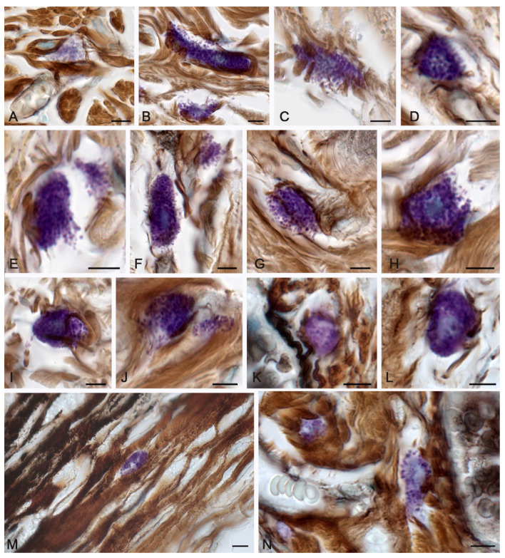
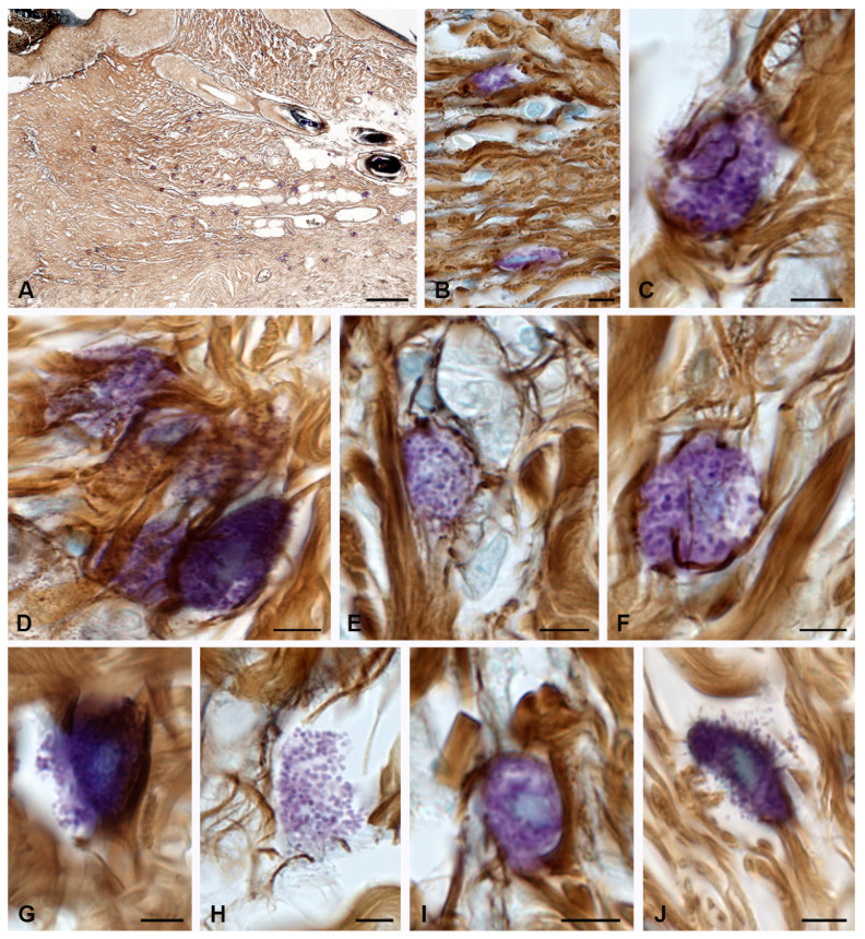
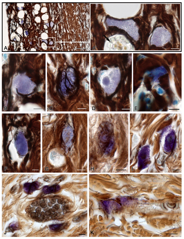
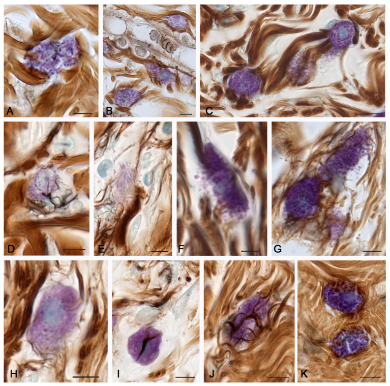
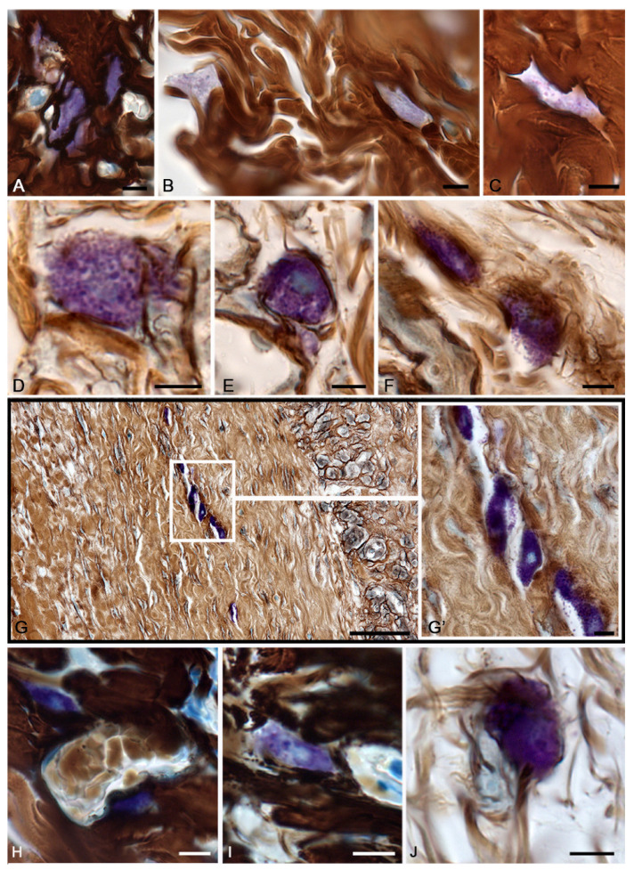
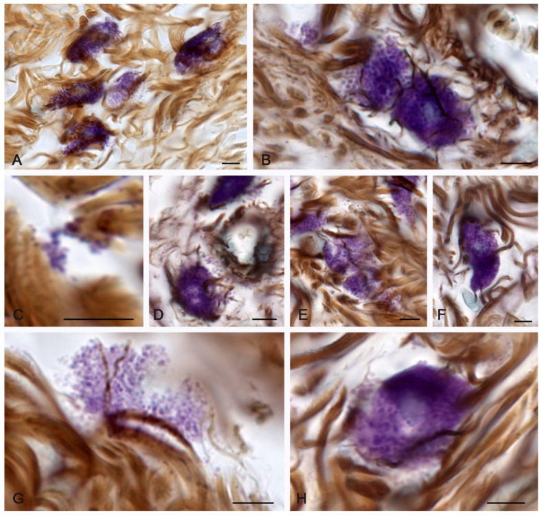
The mechanisms of regeneration for the fibrous component of the connective tissue of the dermis are still insufficiently studied. The aim of this study was to evaluate the effectiveness of the use of molecular hydrogen on the local therapy of a II degree burn wound with the intensification of collagen fibrillogenesis in the skin. We analyzed the involvement of mast cells (MCs) in the regeneration of the collagen fibers of the connective tissue using water with a high content of molecular hydrogen and in a therapeutic ointment for the cell wounds. Thermal burns led to an increase in the skin MC population, accompanied by a systemic rearrangement of the extracellular matrix. The use of molecular hydrogen for the treatment of burn wounds stimulated the regeneration processes by activating the formation of the fibrous component of the dermis, accelerating wound healing. Thus, the intensification of collagen fibrillogenesis was comparable to the effects of a therapeutic ointment. The remodeling of the extracellular matrix correlated with a decrease in the area of damaged skin. Skin regeneration induced by the activation of the secretory activity of MCs may be one of the possible points of implementation of the biological effects of molecular hydrogen in the treatment of burn wounds. Thus, the positive effects of molecular hydrogen on skin repair can be used in clinical practice to increase the effectiveness of therapy after thermal exposure.

Keywords: burn wound; fibrous extracellular matrix; mast cells; molecular hydrogen; regeneration; skin.


Conflict of interest statement

The authors declare no conflict of interest.

Figures

Figure 1
Figure 1
Mast cells of the skin dermis in the post-burn period. The group of spontaneous healing. (AJ)—in 3 days, (KN)—in 7 days of the experiment. (A) A mast cell with a slight degree of metachromasia. (B,C) Active secretion of mast cells with granule localization in the peri-fiber space, with signs of the initial stages of fibrillogenesis (C). (D) Synchronous entry of secretory granules over a large area of MCs into the extracellular matrix. (E) Entry of metachromatic granules into the intercellular substance from different poles of the MCs, formation of a cytoplast with an active secretory potential. (F,G) Granule secretion caused inductive changes in the extracellular matrix, with formation of a fine network of fibers. (H,I) Removal of the granular material was not accompanied by fibrillogenesis. (J) Local secretion of MCs in the direction of collagen fibers, with formation of signs of the initial stages of fibrillogenesis. (K) A perivascular mast cell was involved in the initial stages of collagen fibers formation. (L) Active participation of MCs in fibrillogenesis. (M) Metachromatic secretory material is adjacent to the bundles of dermal collagen fibers of the skin burn surface. (N) Mast cells in the paracrine proximity of the vascular bed. Scale: 5 µm.
Figure 2
Figure 2
Collagen fibers of the skin dermis and mast cells 14 days after burning. (The group of spontaneous healing.) (A) Accumulation of MCs in a certain area of the skin dermis. (B) MCs in the regeneration zone under the burn surface of the skin. (C) Active fiber formation in the mast cell area. (D) MC colocalization in the remodeling cluster of the skin dermis. (E,F) Interaction of MCs and fibroblast (presumably) during collagen fiber formation. (G) Active secretion into the collagen fiber-free area. (H) A non-nuclear metachromatic fragment of the cytoplasm in the area of fibrillogenesis. (I,J) The initial (I) and final (J) stages of collagen fibers formation. Scale: (A)—100 µm, the rest—5 µm.
Figure 3
Figure 3
Mast cells of the skin dermis in the post-burn period under a therapeutic ointment application. (AI)—in 3 days, (JK)—in 7 days of the experiment. (A) Burned surface of the skin with a high content of MCs. (A’) An enlarged fragment of an (A) image. MCs are located next to the microvasculature (arrow). (B) MCs in the lacuna of the dermis after thermal exposure. Secretory granules are distinguished. (C) MCs in the burn surface, with a high level of fibrillogenesis. (D) MCs with altered localization of the secretome in the granules. The nucleus is not determined; a homogeneous formation with preserved properties of metachromasia is detected in the cytoplasm. (E,F) Homogenization of secretory material in MCs in the damaged area of the dermis with weak signs of pericellular fiber formation. (GI) MCs in intact areas of the dermis with different levels of fiber formation intensity. (J) MC group in the peri-venular space of a specific tissue microenvironment, with signs of fibrillogenesis and active degranulation towards the basement membrane of the endothelium. (K) Degranulation of two MCs with the formation of an inductive zone of fibrillogenesis in the local area of secretion. Scale: (A)—50 µm, the rest—5 µm.
Figure 4
Figure 4
Mast cells of the skin dermis 14 days after the onset of the experiment under the therapeutic ointment application. (A) Close MC integration with bundles of collagen fibers. (B) Active MC participation in the formation of the microvascular stroma in the sub-burn zone of the dermis. (C) Joint participation of three MCs in targeted remodeling of the fibrous extracellular matrix of the skin dermis. (D) Initial stages of fibrillogenesis in the peri-burn area of the skin dermis with close MC interaction with fibroblast. (E) Active fibrillogenesis in the region of the MC fragment with low metachromasia. (F,G) Active MC degranulation into the loci of collagen fiber formation induction. (H) The initial stages of the collagen fibrils and fibers formation around the MCs. (I) Low activity of collagen fiber assembly initiation in the area of interaction between MCs and fibroblasts. (J,K) MCs in the zone of dense distribution of the fibrous extracellular matrix with high (J) and low (K) levels of fibrillogenesis. Scale: 5 µm.
Figure 5
Figure 5
Mast cells of the skin dermis in the post-burn period under application of water enriched with molecular hydrogen. (AI)—At 3 days, (JK)—at 7 days of the experiment. (AC) Different variants of MC localization in the damaged skin dermis. There is a homogenization of the secretome and a decrease in the properties of metachromasia (C). (DE) MCs in the peri-burn skin dermis with formation of satellite fragments of the cytoplasm filled with secretome. (F) MC degranulation with collagen fibrillogenesis initiation. (G) Localization of the MC group in the area of the skin dermis restoration, (G’)—highlighted area at a higher magnification. The secretory activity of MCs is noticeable. (HI) MCs in alternative areas of the skin dermis after a burn. Apparently, the MC secretome is partially preserved by this observation period. (J) Active participation of fibroblast-associated MCs in collagen fibrillogenesis. Scale: (G)—50 µm, others—5 µm.
Figure 6
Figure 6
Mast cells of the skin dermis 14 days after the onset of the experiment under application of water enriched with molecular hydrogen. (A,E) Group of actively secreting MCs with the fibrillogenesis loci formation. (B) Two MCs with the reticular fiber formation at the site of adherence to each other. (C) Attachment of MC granules in the extracellular matrix to bundles of collagen fibers with the fibrillogenesis matrix locus formation. (D) Active secretion into the area of the vessel walls of the skin microvasculature. (F) Large MCs without signs of the reticular fiber initiation. (G) Nuclear fragment of the MC cytoplasm with the fibrillogenesis matrix formation. (H) Local MC secretion to the sites of the fibrillogenesis initiation. Scale: 5 µm.

References

    1. Burgess M., Valdera F., Varon D., Kankuri E., Nuutila K. The Immune and Regenerative Response to Burn Injury. Cells. 2022;11:3073. doi: 10.3390/cells11193073. - DOI - PMC - PubMed
    1. Yeganeh P.M., Tahmasebi S., Esmaeilzadeh A. Cellular and biological factors involved in healing wounds and burns and treatment options in tissue engineering. Regen. Med. 2022;17:401–418. doi: 10.2217/rme-2022-0029. - DOI - PubMed
    1. Botting R.A., Haniffa M. The developing immune network in human prenatal skin. Immunology. 2020;160:149–156. doi: 10.1111/imm.13192. - DOI - PMC - PubMed
    1. Davison-Kotler E., Marshall W.S., García-Gareta E. Sources of Collagen for Biomaterials in Skin Wound Healing. Bioengineering. 2019;6:56. doi: 10.3390/bioengineering6030056. - DOI - PMC - PubMed
    1. Plum T., Wang X., Rettel M., Krijgsveld J., Feyerabend T.B., Rodewald H.-R. Human Mast Cell Proteome Reveals Unique Lineage, Putative Functions, and Structural Basis for Cell Ablation. Immunity. 2020;52:404–416.e5. doi: 10.1016/j.immuni.2020.01.012. - DOI - PubMed

LinkOut - more resources