Review
. 2023 Mar 6;15(3):856.
doi: 10.3390/pharmaceutics15030856.

Gene Therapy for Regenerative Medicine

Affiliations
Review

Gene Therapy for Regenerative Medicine

Hossein Hosseinkhani et al. Pharmaceutics. .

Abstract

The development of biological methods over the past decade has stimulated great interest in the possibility to regenerate human tissues. Advances in stem cell research, gene therapy, and tissue engineering have accelerated the technology in tissue and organ regeneration. However, despite significant progress in this area, there are still several technical issues that must be addressed, especially in the clinical use of gene therapy. The aims of gene therapy include utilising cells to produce a suitable protein, silencing over-producing proteins, and genetically modifying and repairing cell functions that may affect disease conditions. While most current gene therapy clinical trials are based on cell- and viral-mediated approaches, non-viral gene transfection agents are emerging as potentially safe and effective in the treatment of a wide variety of genetic and acquired diseases. Gene therapy based on viral vectors may induce pathogenicity and immunogenicity. Therefore, significant efforts are being invested in non-viral vectors to enhance their efficiency to a level comparable to the viral vector. Non-viral technologies consist of plasmid-based expression systems containing a gene encoding, a therapeutic protein, and synthetic gene delivery systems. One possible approach to enhance non-viral vector ability or to be an alternative to viral vectors would be to use tissue engineering technology for regenerative medicine therapy. This review provides a critical view of gene therapy with a major focus on the development of regenerative medicine technologies to control the in vivo location and function of administered genes.

Keywords: biodegradable polymers; gene therapy; nanoparticles; non-viral vectors; regenerative medicine; tissue engineering; viral vectors.


Conflict of interest statement

The authors declare no conflict of interest.
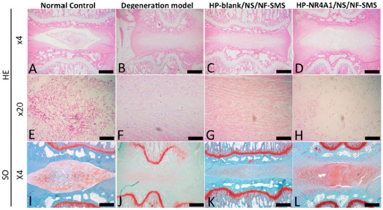
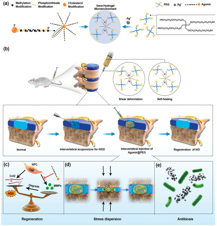
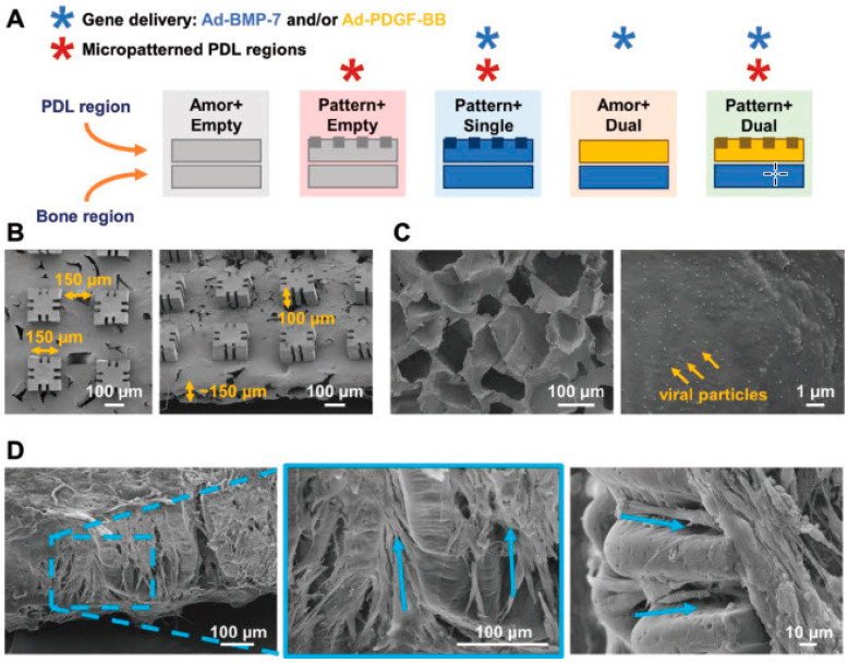
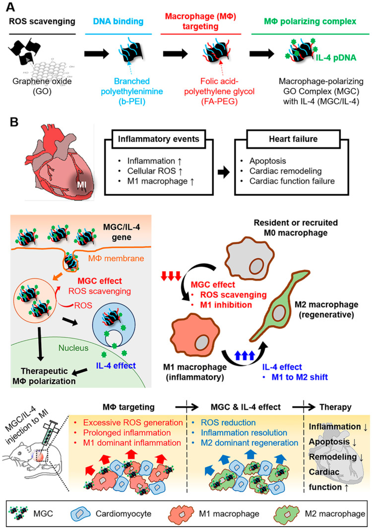
Figures

Figure 1
Figure 1
Anti-tumour efficiency of pHSV-TK-VCMBs with FUS. (A) Quantitative results of HSV-TK expression after using pHSV-TK-VCMBs and FUS treatment (N = 4 per group, compared with untreated group). (B) T2-weighted MRI images of brain tumours at 4, 11, 18, and 25 days after tumour implantation. Rats received no treatment (control group) or treatment on day 7 with: direct injection, pHSV-TK-CMBs, or pHSV-TK-VCMBs. (C) Tumour volume assessed by MRI imaging (N = 6 per group, compared with untreated group). (D) Analysis of animal survival rate (N = 6 per group, compared with untreated group) (* p < 0.5, ** p < 0.01). Reprinted with permission from Ref. [165], Elsevier, copyright 2014.
Figure 2
Figure 2
Timed GDNF protein expression reduces axonal coil formation, improves distal axon outgrowth, and promotes motor neuron survival—results of Experiment 1. (AE) The diameter of ventral roots adjacent to the spinal cord was analysed in ChAT-stained longitudinal sections 12 weeks post-reimplantation. Compared to intact ventral roots, the average diameter increases slightly in CMV-GFP the control group (p < 0.005). (B,C,E) Continuous GDNF protein in the CMV-GDNF and dox-i-GDNF ON groups results in a significant increase in the ventral root diameter. Exposure to time-restricted GDNF expression results in a significant size reduction (D,E), though small isolated nerve coils are occasionally found. ((D) arrow). Constitutive GDNF expression results in densely packed thin fibres growing in a swerving fashion as shown in high magnification ventral root images taken 2 to 8 mm from the implantation site ((H,I) MBP; blue, Neurofilament; green, S100; red). (J) In the ON/OFF group ventral roots were filled with axon profiles displaying a more longitudinal growth pattern. (K) However, coils observed in the ON/OFF group inside the small nerve (shown in (D)), and the axon growth orientation was disrupted similar to the groups with constitutive GDNF expression. (L) Motor neuron survival at 12 weeks improved in all GDNF treated groups. Long distance motor axon outgrowth, quantified in transverse sciatic nerve sections stained for ChAT (M), was significantly increased in the ON/OFF animals. (E) * p < 0.002 versus CMV-GDNF and ON, ** p < 0.0001 versus CMV-GFP. (L) * p < 0.01. (M) # p < 0.008 versus CMV-GFP, p < 0.05 versus CMV-GDNF and ON). Scale bar in A (AD) = 250 µm, K (FK) = 50 µm. Data represent individual animals and are expressed as mean ± SEM. Reprinted from ref. [273] with permission from Oxford University Press, copyright 2019.
Figure 3
Figure 3
Adeno-associated virus-inner ear-Atoh1 (AAV-ie-Atoh1) induces new hair cells (HCs) in vivo with stereocilia. Mice were injected with AAV-ie-Atoh1 (1 × 10¹⁰ genome-containing (particles) (GCs)) at P0, and the cochlea was harvested at P14. (a) Representative confocal projection image of control and AAV-ie-Atoh1 cochlea. Scale bar, 20 μm. (b) Magnification of inner HC (IHC) region of control and AAV-ie-Atoh1 cochlea. White arrows indicate both Sox2-and Myo7a-positive new HCs. Green: Sox2; magenta: Myo7a. Scale bar, 10 μm. (c) Number of Myo7a-positive new HCs per 100 μm in sensory region. Data are shown as mean ± SEM. N = 3. Source data are provided as a Source Data file. (d) Representative confocal image of phalloidin staining of new HCs in AAV-ie-Atoh1 cochlea. Scale bar, 10 μm. (e) Scanning electron microscopy (SEM) images of AAV-ie- and AAV-ie-Atoh1-injected cochlea at apical, middle, and basal regions. Regenerated HC-like cells were artificially coloured magenta. Scale bars, 5 μm (upper panels), 1 μm (lower panels). (f) Representative membrane responses of P14 supporting cells (SCs) to current. The trace shows action potential generation in response to 10 pA injections. N = 5. (g) Same as (f), but for P14 IHCs. The trace shows action potential generation in response to 10 pA injections. The first action potential was generated by 130 pA injection (red arrow). (h) Same as (f), but for P14 new HC injected with AAV-ie-Atoh1 and the first action potential was generated by 20A injection (red arrow). (i) Average responses show significant difference between IHCs and regenerated new HCs. Data are shown as mean ± SEM. p Value is calculated by Student’s t test. *** p < 0.001. N = 5. Reprinted from Ref. [298] with permission from Nature, copyright 2019.
Figure 4
Figure 4
Transplantation of MSCs with CXCR6 gene therapy efficiently regenerated skin in Type I and II diabetic mice. (A) Flow chart representing Type I diabetes model generation. (B) High fasting blood glucose levels (>150 mg/dL) was observed in both Type I and II (db/db) diabetic mice. Higher percent wound closure area in diabetic mice transplanted with MSCs-Cxcr6. (C) Type I Stz-induced C57BL/6J and (D) Type II db/db mice. (E) Higher H&E and Sirius red staining in db/db mice transplanted with MSCs-Cxcr6 depicting an organised layer of dermis with the presence of glands and hair follicles in the regenerated wounds, as compared with control MSCs transplanted groups (E, epidermis; D, dermis; H, Hair follicles; G, sebaceous glands; Ad, adipose layer). Increased co-immunostaining of (F) GFP/CXCR6, (G) GFP/CD31, and (H) GFP/PanCK, suggesting more recruitment, engraftment, neo-vascularisation, and epithelialisation in db/db mice transplanted with MSCs-Cxcr6, as compared with MSC-control. (n = 3 replicates/wound, N = 4–5 mice/group; p < 0.05 as compared with * pLJM1-EGFP control; # diabetic control). Reprinted from Ref. [307] with permission from Elsevier, copyright 2020.
Figure 5
Figure 5
Hematoxylin and eosin (HE) and safranin-O (SO) stained disc images from different experimental groups at four weeks after injection. In the intact discs of the normal control group (A,E), the oval-shaped nucleus pulposus occupied a large volume of the disc space as viewed in the HE-stained midsagittal cross sections. The nucleus pulposus area was stained strongly with safranin-O (I), indicating a high glycosaminoglycan (GAG) content. In the degeneration model group (B,F,J), the disc space collapsed, with evident fibrous tissue invasion. Inhomogeneous fibrous tissue was found in the HP-blank/NS/NF-SMS group (C,G,K), and the nucleus pulposus area was stained almost negatively by safranin O. Although discs in the HPNR4A1 pDNA/NS/NF-SMS group (D,H,L) still displayed some degree of degeneration, therapeutic efficacy was obvious compared to the degeneration model group. The nucleus pulposus area in this group was more similar to the normal control group than other groups and was stained positively with safranin O. Scale bar: (AD,IL) 250 μm, (EH) 100 μm. Reprinted from Ref. [311] with permission from Elsevier, copyright 2017.
Figure 6
Figure 6
Gene-hydrogel microenvironment for regeneration of intervertebral disc degeneration (IVDD). (a) The construction of gene-hydrogel microenvironment. (b) Agomir@PEG was injected into the intervertebral space to construct the gene-hydrogel microenvironment. (ce) The multi-functions provided by the gene-hydrogel microenvironment, matching the regeneration of IVDD. Reprinted from Ref. [322] with permission from WILEY-VCH Verlag GmbH & Co. KGaA, Weinheim, Germany, copyright 2020.
Figure 7
Figure 7
Scaffold design for periodontal tissue neogenesis. (A) Schematic representation of the polymeric scaffolds designed to deliver gene therapy vectors and/or micropatterned surface topography to bony defects. The periodontal ligament (PDL) regions were either patterned or amorphous, made of PLGA/PCL, and seeded with human periodontal ligament cells (hPDLs), except Pattern+Single, which received human gingival fibroblasts (hGFs). The bone regions were amorphous, made of PCL, and seeded with human gingival fibroblasts (hGFs). All regions were CVD-coated to immobilise adenoviral genes for Ad-BMP-7 (blue), Ad-PDGF-BB (yellow), or Ad-empty (gray). (B) SEM images of patterned PDL region, showing the pillar and groove dimensions. (C) SEM images of CVD-coated, PCL, porous base with immobilised adenoviral particles (1012 PN mL−1). (D) SEM images of hPDLs aligned with the micropatterning, 3 d after seeding. Blue arrows indicate the alignment of cells along the grooves embedded within the scaffold pillars, as well as in the interpillar regions. (Ad; adenovirus) Reprinted from Ref. [327] with permission from WILEY-VCH Verlag GmbH & Co. KGaA, Weinheim, copyright 2018.
Figure 8
Figure 8
Schematic illustration of the preparation of the macrophage-polarizing GO complex (MGC)/IL-4, progression of heart failure after MI, and the therapeutic mechanisms of MGC/IL-4 pDNA to treat MI. (A) Stepwise preparation of MGC or MGC/IL-4 pDNA and the role of each chemical conjugation. (B) Progression of heart failure after MI and the therapeutic mechanisms of MGC/IL-4 pDNA in cardiac repair. Reprinted from Ref. [342] with permission from American Chemical Society, copyright 2018.

References

    1. Bessis N., GarciaCozar F.J., Boissier M.C. Immune responses to gene therapy vectors: Influence on vector function and effector mechanisms. Gene Ther. 2004;11((Suppl. S1)):S10–S17. doi: 10.1038/sj.gt.3302364. - DOI - PubMed
    1. Bouard D., Alazard-Dany D., Cosset F.L. Viral vectors: From virology to transgene expression. Br. J. Pharmacol. 2009;157:153–165. doi: 10.1038/bjp.2008.349. - DOI - PMC - PubMed
    1. Baum C., Kustikova O., Modlich U., Li Z., Fehse B. Mutagenesis and oncogenesis by chromosomal insertion of gene transfer vectors. Hum. Gene Ther. 2006;17:253–263. doi: 10.1089/hum.2006.17.253. - DOI - PubMed
    1. Thomas C.E., Ehrhardt A., Kay M.A. Progress and problems with the use of viral vectors for gene therapy. Nat. Rev. Genet. 2003;4:346–358. doi: 10.1038/nrg1066. - DOI - PubMed
    1. Waehler R., Russell S.J., Curiel D.T. Engineering targeted viral vectors for gene therapy. Nat. Rev. Genet. 2007;8:573–587. doi: 10.1038/nrg2141. - DOI - PMC - PubMed

LinkOut - more resources