. 2023 Mar 6;15(3):857.
doi: 10.3390/pharmaceutics15030857.

H2O2-PLA-(Alg)2Ca Hydrogel Enriched in Matrigel® Promotes Diabetic Wound Healing

Affiliations

H2O2-PLA-(Alg)2Ca Hydrogel Enriched in Matrigel® Promotes Diabetic Wound Healing

Alexandra Cătălina Bîrcă et al. Pharmaceutics. .

Abstract

Hydrogel-based dressings exhibit suitable features for successful wound healing, including flexibility, high water-vapor permeability and moisture retention, and exudate absorption capacity. Moreover, enriching the hydrogel matrix with additional therapeutic components has the potential to generate synergistic results. Thus, the present study centered on diabetic wound healing using a Matrigel-enriched alginate hydrogel embedded with polylactic acid (PLA) microspheres containing hydrogen peroxide (H2O2). The synthesis and physicochemical characterization of the samples, performed to evidence their compositional and microstructural features, swelling, and oxygen-entrapping capacity, were reported. For investigating the three-fold goal of the designed dressings (i.e., releasing oxygen at the wound site and maintaining a moist environment for faster healing, ensuring the absorption of a significant amount of exudate, and providing biocompatibility), in vivo biological tests on wounds of diabetic mice were approached. Evaluating multiple aspects during the healing process, the obtained composite material proved its efficiency for wound dressing applications by accelerating wound healing and promoting angiogenesis in diabetic skin injuries.

Keywords: Matrigel; alginate-hydrogel-based dressing; oxygen-enriched polylactic acid microspheres; skin; wound healing.


Conflict of interest statement

The authors declare no conflict of interest.

Figures

Figure 1
Figure 1
The surgical procedure used on the murine model. (A) Execution of the skin defect. (B) Placement of the silicone rings. (C) Placement of hydrogels.
Figure 2
Figure 2
The FT-IR spectra of HG, Oms, and HG_OMs.
Figure 3
Figure 3
SEM images of OMs (a,b).
Figure 4
Figure 4
SEM images of HG control sample (a,b) and pore size distribution (c).
Figure 5
Figure 5
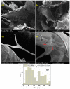
SEM images of HG_OMs (ad) and pore size distribution (e).
Figure 6
Figure 6
Swelling rate (a) and degradation (b) of HG and HG_OMs.
Figure 7
Figure 7
Gross morphology of wounds at different time points after surgery. (Right) Wound closures were observed on days 3 and 7 after the wound injury. (a) Full-thickness skin defect (control); (b) alginate hydrogel dressing (HG); (c) oxygen-releasing alginate hydrogel containing H2O2-PLA microspheres (HG_OMs); (d) oxygen-releasing hydrogel, containing H2O2-PLA microspheres and 4.44 μg/mg (HG_OMs_MG). (Left) Wound closure size (cm2).
Figure 8
Figure 8
The histological aspect of the full-thickness skin defect at 3 days (H&E stain). Symbols: dotted line—separates healthy skin from the defect; GT—granulation tissue; *—edema; circle—neutrophils, K—keratin.
Figure 9
Figure 9
The histological aspect of the skin defect filled with HG at 3 days (H&E stain). Symbols: dotted line—separates healthy skin from the defect; GT—granulation tissue; circle—neutrophils.
Figure 10
Figure 10
The histological aspect of the skin defect filled with HG_OMs at 3 days (H&E stain). Symbols: GT—granulation tissue.
Figure 11
Figure 11
The histological aspect of the skin defect filled with HG-OMs_-MG at 3 days (H&E stain). Symbols: GT—granulation tissue; circle—extended vascular area; arrow—neutrophil extravasation.
Figure 12
Figure 12
The histological aspect of the full-thickness skin defect at 7 days (H&E stain). Symbols: dotted line—separates healthy skin from the defect; circle, arrow—new capillaries.
Figure 13
Figure 13
The histological aspect of the skin defect filled with HG at 7 days (H&E stain). Symbols: NE—new epidermis; arrow—epithelial cells in the dermis.
Figure 14
Figure 14
The histological aspect of the skin defect filled with HG-OMs at 7 days (H&E stain). Symbols: dotted line—separates healthy skin from the defect; NE—new epidermis; arrow—new capillaries.
Figure 15
Figure 15
The histological aspect of the skin defect filled with HG-OMs-MG at 7 days (H&E stain). Symbols: dotted line—separates healthy skin from the defect; NE—new epidermis; circle—the proliferation of capillaries in a mature dermis; arrow—new capillaries.
Figure 16
Figure 16
The histological aspect of the keratinization process during the healing process of the epidermis (Dane stain).

References

    1. Zhang X., Kang X., Jin L., Bai J., Liu W., Wang Z. Stimulation of wound healing using bioinspired hydrogels with basic fibroblast growth factor (bFGF) Int. J. Nanomed. 2018;13:3897. doi: 10.2147/IJN.S168998. - DOI - PMC - PubMed
    1. Guebitz G.M., Nyanhongo G.S. Enzymes as Green Catalysts and Interactive Biomolecules in Wound Dressing Hydrogels. Trends Biotechnol. 2018;36:1040–1053. doi: 10.1016/j.tibtech.2018.05.006. - DOI - PubMed
    1. Atma Y. Synthesis and Application of Fish Gelatin for Hydrogels/Composite Hydrogels: A Review. Biointerface Res. Appl. Chem. 2022;12:3966–3976. doi: 10.33263/briac123.39663976. - DOI
    1. Sun M., Zhu C., Long J., Lu C., Pan X., Wu C. PLGA microsphere-based composite hydrogel for dual delivery of ciprofloxacin and ginsenoside Rh2 to treat Staphylococcus aureus-induced skin infections. Drug Deliv. 2020;27:632–641. doi: 10.1080/10717544.2020.1756985. - DOI - PMC - PubMed
    1. Suo H., Hussain M., Wang H., Zhou N., Tao J., Jiang H., Zhu J. Injectable and pH-Sensitive Hyaluronic Acid-Based Hydrogels with On-Demand Release of Antimicrobial Peptides for Infected Wound Healing. Biomacromolecules. 2021;22:3049–3059. doi: 10.1021/acs.biomac.1c00502. - DOI - PubMed

LinkOut - more resources