. 2023 Mar 15;15(3):953.
doi: 10.3390/pharmaceutics15030953.

Clinically Expired Platelet Concentrates as a Source of Extracellular Vesicles for Targeted Anti-Cancer Drug Delivery

Affiliations

Clinically Expired Platelet Concentrates as a Source of Extracellular Vesicles for Targeted Anti-Cancer Drug Delivery

Ana Meliciano et al. Pharmaceutics. .

Abstract

The short shelf life of platelet concentrates (PC) of up to 5-7 days leads to higher wastage due to expiry. To address this massive financial burden on the healthcare system, alternative applications for expired PC have emerged in recent years. Engineered nanocarriers functionalized with platelet membranes have shown excellent targeting abilities for tumor cells owing to their platelet membrane proteins. Nevertheless, synthetic drug delivery strategies have significant drawbacks that platelet-derived extracellular vesicles (pEV) can overcome. We investigated, for the first time, the use of pEV as a carrier of the anti-breast cancer drug paclitaxel, considering it as an appealing alternative to improve the therapeutic potential of expired PC. The pEV released during PC storage showed a typical EV size distribution profile (100-300 nm) with a cup-shaped morphology. Paclitaxel-loaded pEV showed significant anti-cancer effects in vitro, as demonstrated by their anti-migratory (>30%), anti-angiogenic (>30%), and anti-invasive (>70%) properties in distinct cells found in the breast tumor microenvironment. We provide evidence for a novel application for expired PC by suggesting that the field of tumor treatment research may be broadened by the use of natural carriers.

Keywords: anti-angiogenic potential; drug delivery system; expired platelet concentrates; human breast cancer cell line; paclitaxel; platelet-derived extracellular vesicles.


Conflict of interest statement

The authors declare no conflict of interest.

Figures

Figure 1
Figure 1
Characterization of platelets and isolated platelet-derived extracellular vesicles (pEV). (A) Schematic overview of the density gradient ultracentrifugation (DGUC) protocol used to isolate pEV from expired platelet concentrates (PC). (B) Expression of platelet activation marker CD62p in basal and thrombin-stimulated platelets (n = 3; two-tailed unpaired t-test, *** p < 0.001). (C) Representative transmission electron microscopy (TEM) micrographs of platelets from the expired PC (activated platelets are indicated by black arrows and resting platelets by red arrows). Scale bars: 1 μm. (D) Western blot analysis of specific EV markers (tetraspanins CD63 (glycosylated form) and CD9, and cytosolic protein flotillin-2), platelet-specific marker (CD41), and non-EV markers [apolipoprotein (ApoA1) and argonaute 2 (Ago2)] in pooled fractions and platelet lysate (PLTS). Molecular weight (MW) markers are indicated. (E) Representative TEM images of negatively stained 8–9 fractions, enriched in cup-shaped pEV (black arrows). Higher magnification image detailing the morphology of pEV. Scale bars: 1 µm and 200 nm (high magnification). (F) Representative size distribution profiles of 8–9 pooled pEV-fractions analyzed using nanoparticle tracking analysis. Size distribution is represented as mean (black continuous line) ± standard deviation (shaded area).
Figure 2
Figure 2
Cellular uptake of PKH26-pEV into HUVEC, MDA-MB-231, and BT474 cells. (A) Representative immunofluorescence images of PKH26-pEV uptake. Cells were incubated with PKH26-pEV (Red, at a density of 6000 pEV/cell) for 24 h. HUVEC, MDA-MB-231, and BT474 cells were stained for CD31, EGFR, HER2 (green), respectively, and nuclei (DAPI, blue). Scale bars: 50 μm. (B) PKH26-pEV positive cells were quantitatively measured by flow cytometry 24 and 48 h after PKH26-pEV treatment. Blue filled histograms show the negative control population (cells incubated with DPBS-PKH26), and red filled histograms represent the PKH26-pEV population. The x-axis represents detection by the DsRed-A filter and the y-axis represents pEV counts.
Figure 3
Figure 3
Loading of paclitaxel into pEV by direct incubation and characterization of PTX-pEV. (A) Schematic workflow of PTX loading protocol in pEV by direct incubation. (B) Representative negative staining TEM images of DMSO-pEV, and PTX-pEV. Scale bars: 500 nm and 200 nm (high magnification). (C) Mode and (D) mean particle size (nm) of pEV, DMSO-pEV, and PTX-pEV, as measured by NTA (n = 3). n represents biologically independent replicates. Data are represented as mean ± S.D. One-way ANOVA followed by Tukey’s multiple comparison test, * p < 0.05, n.s., not significant.
Figure 4
Figure 4
Effect of PTX-pEV on HUVEC angiogenesis. (A) Representative images of HUVEC tube formation assay cultured on growth factor-reduced matrigel treated with pEV, DMSO-pEV (treatment vehicle), and PTX-pEV at a density of 6000 pEV/cell. Scale bars: 200 μm. (B) Total segments length (µm), and (C) number of nodes after 8 h of treatment (n = 3). n represents biologically independent replicates. Data are represented as mean ± S.D. One-way ANOVA followed by Tukey’s multiple comparison test, * p < 0.05, ** p < 0.01, n.s., not significant.
Figure 5
Figure 5
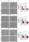
Effects of PTX-pEV on HUVEC, MDA-MB-23, and BT474 cell migration. (A,C,E) Representative images at the initial time (0 h) and wound healing at 16 h for HUVEC, 24 h for MDA-MB-231, and 48 h for BT474 cells incubated PTX-pEV (6000 PTX-pEV/cell). As a control, pEV were used at a density of 6000 pEV/cell. Scale bars: 200 µm. (B,D,F) Quantitative analysis of the wound closure percentage for cells treated with pEV, DMSO-pEV (treatment vehicle), PTX-pEV, free DMSO and free PTX (0.5 μM and 1 µM) at 16 h (B), 24 h (D), and 48 h (F); (n = 3). n represents biologically independent replicates. Data are represented as mean ± S.D. One-way ANOVA followed by Tukey’s multiple comparison test, * p < 0.05, ** p < 0.01, n.s. not significant.
Figure 6
Figure 6
Effect of PTX-pEV on MDA-MB-231 cell invasiveness. (A) Representative images at the initial time (0 h) and wound healing at 24 h and 48 h for MDA-MB-231 breast cancer cells incubated with PTX-pEV (6000 PTX-pEV/cell). As controls, pEV and DMSO-pEV (treatment vehicle) were used at a density of 6000 pEV/cell. Scale bars: 200 μm. (B) Quantitative analysis of the relative wound density percentage after 48 h of treatment for cells treated with pEV, DMSO-pEV, PTX-pEV, free DMSO, and free PTX (0.5 μM and 1 µM); (n = 3). n represents biologically independent replicates. Data are represented as mean ± S.D. One-way ANOVA followed by Tukey’s multiple comparison test, * p < 0.05, **** p < 0.0001.

References

    1. World Health Organization. WHO Model List of Essential Medicines: 20th List. 2017. [(accessed on 2 November 2022)]. Available online: https://apps.who.int/iris/handle/10665/273826.
    1. Hofmann A., Ozawa S., Shander A. Activity-based cost of platelet transfusions in medical and surgical inpatients at a US hospital. Vox Sang. 2021;116:998–1004. doi: 10.1111/vox.13095. - DOI - PubMed
    1. Estcourt L.J. Why has demand for platelet components increased? A review. Transfus. Med. 2014;24:260–268. doi: 10.1111/tme.12155. - DOI - PubMed
    1. Burnouf T., Goubran H.A., Seghatchian J. Multifaceted regenerative lives of expired platelets in the second decade of the 21st century. Transfus. Apher. Sci. 2014;51:107–112. doi: 10.1016/j.transci.2014.08.006. - DOI - PubMed
    1. Prakash S., Thakur A. Platelet concentrates: Past, present and future. J. Maxillofac. Oral. Surg. 2011;10:45–49. doi: 10.1007/s12663-011-0182-4. - DOI - PMC - PubMed

LinkOut - more resources