. 2023 Mar 10;12(6):1263.
doi: 10.3390/plants12061263.

Comparative Nectary Morphology across Cleomaceae (Brassicales)

Affiliations

Comparative Nectary Morphology across Cleomaceae (Brassicales)

Brandi Zenchyzen et al. Plants (Basel). .

Abstract

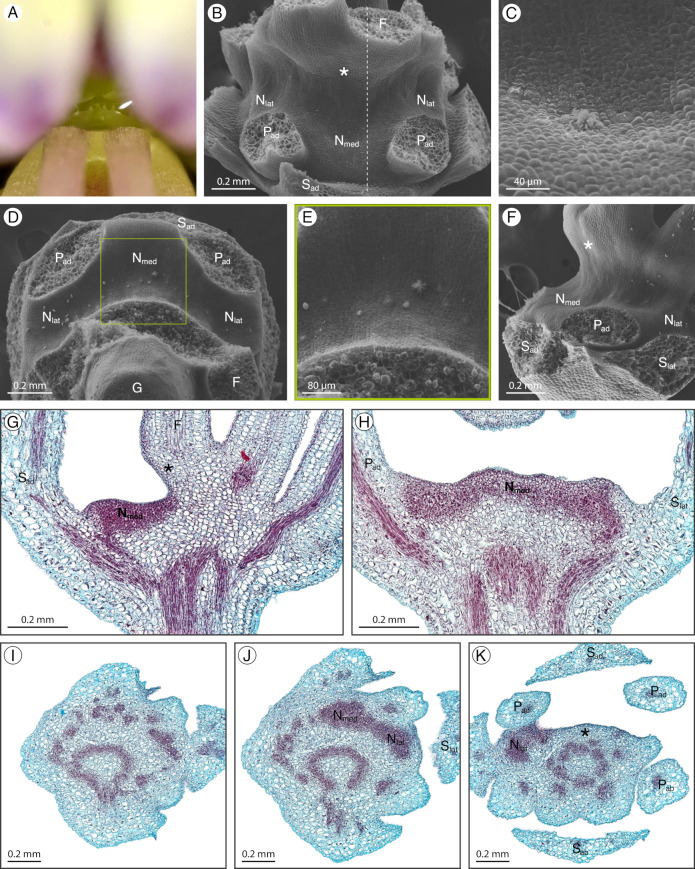
Floral nectaries have evolved multiple times and rapidly diversified with the adaptive radiation of animal pollinators. As such, floral nectaries exhibit extraordinary variation in location, size, shape, and secretory mechanism. Despite the intricate ties to pollinator interactions, floral nectaries are often overlooked in morphological and developmental studies. As Cleomaceae exhibits substantial floral diversity, our objective was to describe and compare floral nectaries between and within genera. Floral nectary morphology was assessed through scanning electron microscopy and histology across three developmental stages of nine Cleomaceae species including representatives for seven genera. A modified fast green and safranin O staining protocol was used to yield vibrant sections without highly hazardous chemicals. Cleomaceae floral nectaries are most commonly receptacular, located between the perianth and stamens. The floral nectaries are supplied by vasculature, often contain nectary parenchyma, and have nectarostomata. Despite the shared location, components, and secretory mechanism, the floral nectaries display dramatic diversity in size and shape, ranging from adaxial protrusions or concavities to annular disks. Our data reveal substantive lability in form with both adaxial and annular floral nectaries interspersed across Cleomaceae. Floral nectaries contribute to the vast morphological diversity of Cleomaceae flowers and so are valuable for taxonomic descriptions. Though Cleomaceae floral nectaries are often derived from the receptacle and receptacular nectaries are common across flowering plants, the role of the receptacle in floral evolution and diversification is overlooked and warrants further exploration.

Keywords: Cleomaceae; evo-devo; floral nectary; floral rewards; floral structure; morphology; nectar secretion; nectarostomata.


Conflict of interest statement

The authors declare no conflict of interest.

Figures

Figure 2
Figure 2
Arivela viscosa nectary (A) photograph, (BF) scanning electron micrographs, and (GK) fast green and safranin O-stained sections. (A) Apical view of the nectary. (B) Adaxial view of the nectary. (CE) Adaxial view close-ups: (C,D) medial nectary lobe, corresponding to the left and right boxes in (B), respectively, and (E) lateral nectary lobe. (F) Apical view of the nectary. (G,H) Longitudinal sections (frontal plane), corresponding to the bottom and top dashed lines in (F), respectively. (IK) Transverse sections of the nectary from proximal to distal positioning. All images are of anthetic stage specimens. F: filament; Nlat: lateral nectary lobe; Nmed: medial nectary lobe; Pab: abaxial petal; Pad: adaxial petal; Sab: abaxial sepal; Sad: adaxial sepal; Slat: lateral sepal.
Figure 3
Figure 3
Cleome amblyocarpa nectary (A) photograph, (BF) scanning electron micrographs, and (GL) fast green and safranin O-stained sections. (A) Apical view of the nectary. (B) Adaxial view of the nectary. (C) Adaxial view close-up of the medial nectary lobe, corresponding to the box in (B). (D) Apical view of the nectary. (E,F) Apical view close-ups of the lateral nectary lobes, corresponding to the left and right boxes in (D), respectively. (GI) Longitudinal sections (sagittal plane) of the nectary, corresponding to the dashed lines in (B) from right to left, respectively. (JL) Transverse sections of the intermediate stage nectary from proximal to distal positioning. All images are of anthetic stage specimens unless indicated otherwise. F: filament; G: gynophore; Nlat: lateral nectary lobe; Nmed: medial nectary lobe; Pab: abaxial petal; Pad: adaxial petal; Sab: abaxial sepal; Sad: adaxial sepal.
Figure 4
Figure 4
Cleome violacea nectary (A) photograph, (BG) scanning electron micrographs, and (HL) fast green and safranin O-stained sections. (A) Apical view of the nectary. (B) Adaxial view of the nectary. (C) Adaxial view close-up of the medial and lateral nectary lobes, corresponding to the box in (B). (D) Apical view of the nectary. (E) Lateral view of the nectary. (F) Lateral view close-up of the nectary. (G) Close-up of nectarostomata. (H,I) Longitudinal sections (sagittal plane) of the nectary, corresponding to the right and left dashed lines in (B), respectively: (H) intermediate stage nectary, and (I) anthetic stage nectary. (JL) Transverse sections: (J) intermediate stage nectary, (K,L) anthetic stage nectary from proximal to distal positioning. All images are of anthetic stage specimens unless indicated otherwise. F: filament; G: gynophore; Nlat: lateral nectary lobe; Nmed: medial nectary lobe; Pab: abaxial petal; Pad: adaxial petal; Sad: adaxial sepal; Slat: lateral sepal.
Figure 5
Figure 5
Gynandropsis gynandra nectary (A) photograph, (BH) scanning electron micrographs, and (IL) fast green/safranin O-stained sections. (A) Apical view of the nectary. (B) Side view of the nectary. (C) Apical view of the nectary. (D) Apical view close-up of the bud stage nectary. (E,F) Close-up of nectarostomata. (G,H) Close-up of the nectary. (I) Longitudinal section of the intermediate stage nectary. (JL) Transverse sections of the nectary, corresponding to the dashed lines in (B) from proximal to distal, respectively. All images are of anthetic stage specimens unless indicated otherwise. A: androgynophore; F: filament; N: nectary; P: petal; S: sepal.
Figure 6
Figure 6
Melidiscus giganteus nectary (A) photograph, (BF) scanning electron micrographs, and (GL) fast green and safranin O-stained sections. (A) Apical view of the nectary. (B) Adaxial view of the nectary. (C) Apical view of the nectary. (D,E) Adaxial view close-ups, corresponding to the bottom and top boxes in (B), respectively. (F) Apical view close-up, corresponding to the box in (C). (G,H) Longitudinal sections (frontal plane): (G) lateral nectary lobes and (H) abaxial side of nectary. (IL) Transverse sections of the nectary, corresponding to the dashed lines in (B) from proximal to distal, respectively. All images are of anthetic stage specimens. F: filament; G: gynophore; Nlat: lateral nectary lobe; Nmed: medial nectary lobe; Pab: abaxial petal; Pad: adaxial petal; Sab: abaxial sepal; Sab: abaxial sepal; Slat: lateral sepal.
Figure 7
Figure 7
Polanisia dodecandra nectary (A) photograph, (BF) scanning electron micrographs, and (GL) fast green and safranin O-stained sections. (A) Apical view of the nectary. (B) Adaxial view of the nectary. (C) Apical view of the nectary. (DF) Adaxial view close-ups: (D) bud stage nectary, (E) anthetic stage nectary, corresponding to the box in (B), and (F) anthetic stage nectarostomata. (G) Longitudinal section (frontal plane) of the intermediate stage nectary. (H,I) Longitudinal section (sagittal plane): (H) intermediate stage nectary and (I) anthetic stage nectary. (JL) Transverse sections of the nectary from proximal to distal positioning. All images are of anthetic stage specimens unless indicated otherwise. F: filament; G: gynophore; N: nectary; Pad: adaxial petal; Sad: adaxial sepal.
Figure 8
Figure 8
Sieruela hirta nectary (A) photograph, (BF) scanning electron micrographs, and (GK) fast green and safranin O-stained sections. (A) Apical view of the nectary. (B) Adaxial view of the nectary. (C) Adaxial view close-up of the nectary, corresponding to the box in (B). (D) Apical view of the nectary. (E) Lateral view of the nectary. (F) Lateral view close-up of the nectary, corresponding to the box in (E). (G) Longitudinal section (sagittal plane) of the intermediate stage nectary, corresponding to the dashed line in (D). (HK) Transverse sections of the nectary from proximal to distal orientation. All images are of anthetic stage specimens unless indicated otherwise. Basal fusion of adaxial filaments is indicated with an asterisk. F: filament; Nlat: lateral nectary lobe; Nmed: medial nectary lobe; Pab: abaxial petal; Pad: adaxial petal; Sad: adaxial sepal.
Figure 9
Figure 9
Sieruela rutidosperma nectary (A) photograph, (BF) scanning electron micrographs, and (GK) fast green and safranin O-stained sections. (A) Apical view of the nectary. (B) Adaxial view of the nectary. (C) Adaxial view close-up of the medial nectary lobe. (D) Apical view of the nectary. (E) Apical view close-up of the nectary, corresponding to the box in (D). (F) Lateral view of the nectary. (G) Longitudinal section (sagittal plane) of the intermediate stage nectary, corresponding to the dashed line in (B). (H) Longitudinal section (frontal plane) of the nectary. (IK) Transverse section of the nectary from proximal to distal positioning. All images are of anthetic stage specimens unless indicated otherwise. Basal fusion of adaxial filaments is indicated with an asterisk. F: filament; G: gynophore; Nlat: lateral nectary lobe; Nmed: medial nectary lobe; Pab: abaxial petal; Pad: adaxial petal; Sab: abaxial sepal; Sad: adaxial sepal; Slat: lateral sepal.
Figure 10
Figure 10
Tarenaya houtteana nectary (A) photograph, (BH) scanning electron micrographs, and (IM) fast green and safranin O-stained sections. (A) Apical view of the nectary. (B) Adaxial view of the nectary. (C) Apical view of the nectary. (D) Adaxial view close-up of the nectary, corresponding to the box in (B). (E) Apical view close-up of the nectary, corresponding to the box in (C). (FH) Close-up of nectarostomata. (I) Longitudinal section (sagittal plane) of the bud stage nectary. (J) Longitudinal section (frontal-oblique plane) of the intermediate stage nectary. (KM) Transverse sections of the nectary from proximal to distal positioning. All images are of anthetic stage specimens unless indicated otherwise. F: filament; G: gynophore; Nlat: lateral nectary lobe; Nmed: medial nectary lobe; Pab: abaxial petal; Pad: adaxial petal; Sab: abaxial sepal; Sad: adaxial sepal; Slat: lateral sepal.
Figure 11
Figure 11
Nectarostomata of the nine Cleomaceae species. (A) Arivela viscosa. (B) Cleome amblyocarpa. (C) Cleome violacea. (D) Gynandropsis gynandra. (E) Melidiscus giganteus. (F) Polanisia dodecandra. (G) Sieruela hirta. (H) Sieruela rutidosperma. (I) Tarenaya houtteana. All images are of anthetic stage specimens, except C. amblyocarpa (intermediate stage) and G. gynandra (bud stage). Scale bar represents 10 μm.
Figure 12
Figure 12
Cleomaceae phylogeny and floral nectaries of the nine species grouped by shape and position. (A) Cleomaceae phylogenetic tree derived from that of Patchell et al. (2014) and Bayat et al. (2018) [7,22] with clades sampled here bolded. (B) Annular nectaries. (C) Protruding adaxial nectaries. (D) Slightly convex adaxial nectary. (E) Concave adaxial nectaries. Illustrations depict the apical view of the nectary with stippling to represent more basal or concave regions. Nectary size varies between species; illustrations are not to the same scale.
Figure 1
Figure 1
Anthetic flower with nectary inset of the nine Cleomaceae species. (A) Arivela viscosa. (B) Cleome amblyocarpa. (C) Cleome violacea. (D) Gynandropsis gynandra. (E) Melidiscus giganteus. (F) Polanisia dodecandra. (G) Sieruela hirta. (H) Sieruela rutidosperma. (I) Tarenaya houtteana. The scale bar represents 0.25 cm in all images to illustrate the drastic variation in flower size.

References

    1. Willmer P. Pollination and Floral Ecology. Princeton University Press; Princeton, NJ, USA: 2011.
    1. Roy R., Schmitt A.J., Thomas J.B., Carter C.J. Review: Nectar biology: From molecules to ecosystems. Plant Sci. 2017;262:148–164. doi: 10.1016/j.plantsci.2017.04.012. - DOI - PubMed
    1. Slavković F., Dogimont C., Morin H., Boualem A., Bendahmane A. The genetic control of nectary development. Trends Plant Sci. 2021;26:260–271. doi: 10.1016/j.tplants.2020.11.002. - DOI - PubMed
    1. Nicolson S.W., Nepi M., Pacini E. Nectaries and Nectar. Springer; Dordrecht, The Netherlands: 2007.
    1. Liao I.T., Hileman L.C., Roy R. On the horizon for nectar-related research. Am. J. Bot. 2021;108:2326–2330. doi: 10.1002/ajb2.1767. - DOI - PubMed