. 2023 May;6(5):e1810.
doi: 10.1002/cnr2.1810. Epub 2023 Mar 28.

Utilization of a cell-penetrating peptide-adaptor for delivery of human papillomavirus protein E2 into cervical cancer cells to arrest cell growth and promote cell death

Affiliations

Utilization of a cell-penetrating peptide-adaptor for delivery of human papillomavirus protein E2 into cervical cancer cells to arrest cell growth and promote cell death

Julia C LeCher et al. Cancer Rep (Hoboken). 2023 May.

Abstract

Background: Human papillomavirus (HPV) is the causative agent of nearly all forms of cervical cancer, which can arise upon viral integration into the host genome and concurrent loss of viral regulatory gene E2. Gene-based delivery approaches show that E2 reintroduction reduces proliferative capacity and promotes apoptosis in vitro.

Aims: This work explored if our calcium-dependent protein-based delivery system, TAT-CaM, could deliver functional E2 protein directly into cervical cancer cells to limit proliferative capacity and induce cell death.

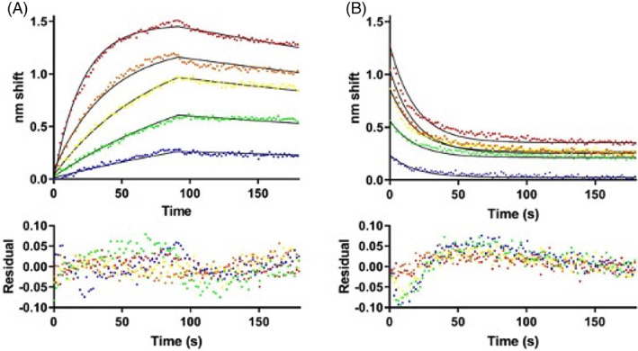
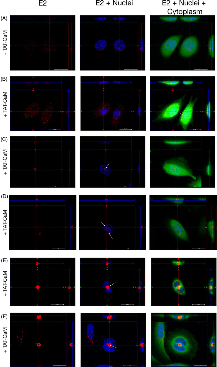
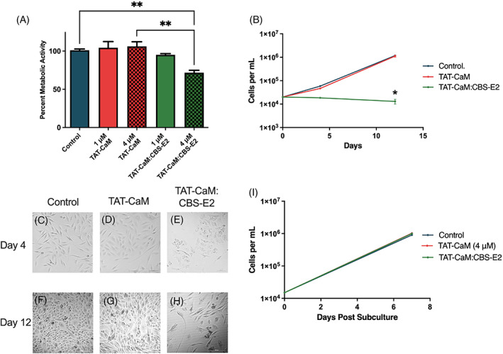
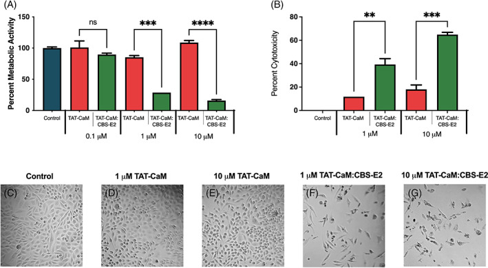
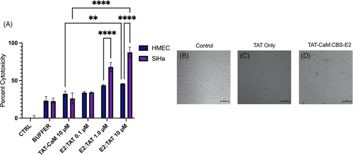
Materials and results: TAT-CaM and the HPV16 E2 protein containing a CaM-binding sequence (CBS-E2) were expressed and purified from Escherichia coli. Calcium-dependent binding kinetics were verified by biolayer interferometry. Equimolar TAT-CaM:CBS-E2 constructs were delivered into the HPV16+ SiHa cell line and uptake verified by confocal microscopy. Proliferative capacity was measured by MTS assay and cell death was measured by release of lactate dehydrogenase. As a control, human microvascular cells (HMECs) were used. As expected, TAT-CaM bound CBS-E2 with high affinity in the presence of calcium and rapidly disassociated upon its removal. After introduction by TAT-CaM, fluorescently labeled CBS-E2 was detected in cellular interiors by orthogonal projections taken at the depth of the nucleus. In dividing cells, E2 relocalized to regions associated with the mitotic spindle. Cells receiving a daily dose of CBS-E2 for 4 days showed a significant reduction in metabolic activity at low doses and increased cell death at high doses compared to controls. This phenotype was retained for 7 days with no further treatments. When subcultured on day 12, treated cells regained their proliferative capacity.

Conclusions: Using the TAT-CaM platform, bioactive E2 protein was delivered into living cervical cancer cells, inducing senescence and cell death in a time- and dose-dependent manner. These results suggest that this nucleic acid and virus-free delivery method could be harnessed to develop novel, effective protein therapeutics.

Keywords: E2; E6; E7; HPV-16; cell-penetrating peptides; cervical cancer.


Conflict of interest statement

The authors have stated explicitly that there are no conflicts of interest in connection with this article.

Figures

FIGURE 1
FIGURE 1
Biolayer interferometry analysis of TAT‐CaM binding to CBS‐E2. (A) Association‐then‐dissociation experiment in which ligand TAT‐CaM was exposed to varying concentrations of CBS‐E2 prior to movement to buffer only at 90s (red, 1000 nM; orange, 500 nM; yellow, 250 nM, green, 125 nM, blue 63 nM). Data points are individual instrument readings. Lines represent best fits to a global single‐state model. Residuals are shown below. (B) The same samples after dissociation were moved to buffer containing 10 mM EDTA for monitoring of dissociation in the absence of Ca2+. Fits are to a global single‐state exponential decay model. Residuals indicate some non‐ideality in the model, likely due to rapid dissociation prior to the first reading (see discussion).
FIGURE 2
FIGURE 2
Delivery of CBS‐E2 into living cervical cancer cells via the TAT‐CaM adaptor. Cervical cancer cells were incubated with fluorescently labeled CBS‐E2 cargo (Red) in the absence (A) or presence (B–F) of equimolar TAT‐CaM for 1 h. Cells were counterstained with NucBlue (nuclei; blue) and Cytotracker (cytoplasm; green). Images were generated on an inverted Zeiss LSM700 Confocal Microscope with Z‐stack projections. Shown at the top and right of each image are orthogonal projections taken at the depth of the nucleus. (A, B) Visualization of E2 in SiHas in asynchronous populations. (C–F) Visualization of CBS‐E2 in mitotically active cells. White arrows indicate redistribution and clustering of CBS‐E2 to regions of the cell typically associated with the mitotic spindle apparatus.
FIGURE 3
FIGURE 3
CBS‐E2 delivery induces reversible inhibition of cell growth in cervical cancer cells. Cells were seeded at 2.5 × 104 per well and treated once daily for 3 days with either 1 or 4 μM CBS‐E2 in the presence of equimolar amounts of TAT‐CaM. As a control, cells were either left untreated (negative control) or treated with TAT‐CaM only (experimental control). (A) MTS assay to assess cellular metabolic activity on day 4. A reduction in metabolic activity was tested for by One‐way ANOVA with Dunnet's correction for multiple comparisons **control to CBS‐E2 4 μM p = .004, **TAT‐CaM 4 μM to CBS‐E2 4 μM p = .001. n = 9 for all groups; shown SEM. (B) On day 4 and day 12, cells were collected and counted on a hemocytometer. Data were analyzed by Two‐way ANOVA with Dunnet's correction for multiple comparisons to assess the effect of both time and treatment. p‐value indicates comparison between the TAT‐CaM and the TAT‐CaM:CBS‐E2 group *p = .02. n = 4 for all groups; shown SEM. (C–H) Micrographs of cells from each treatment group on day 4 and day 12 post‐treatment. Images are from the 4 μM treatments. (I) On day 12, cells were collected and reseeded at equal density and cultured for an additional week after which they were collected and counted on a hemocytometer. n = 4 for all groups.
FIGURE 4
FIGURE 4
CBS‐E2 induces cell death in cervical cancer cells. Cells were seeded at 2.5 × 103 and treated once daily for 3 days with either 1, 3 or 10 μM CBS‐E2 in the presence of equimolar amounts of TAT‐CaM. As a control, cells were either left untreated (negative control) or treated with TAT‐CaM only (experimental control). (A) MTS assay to assess cellular metabolic activity on day 4. A reduction in metabolic activity was tested for by One‐way ANOVA with Dunnet's correction for multiple comparisons. p‐value indicates comparison between the TAT‐CaM and the TAT‐CaM:CBS‐E2 group at same concentrations ****p < .001. n = 9 for all groups; shown SEM. (B) LDH leakage assay to assess cytotoxicity on day 4. Percent cytotoxicity was tested for by one‐way ANOVA with Dunnet's correction for multiple comparisons. p‐value indicates comparison between the TAT‐CaM and the TAT‐CaM:CBS‐E2 group at same concentration ***p = .007, ***p = .0006, n = 9 for all groups; shown SEM. (C–G) Micrographs of cells from each treatment group taken on day 4.
FIGURE 5
FIGURE 5
CBS‐E2 does not induce cell death in human microvascular endothelial cells. SiHa and HMEC cells were seeded at 2.5 × 103 and treated once daily for 3 days with 10 μM CBS‐E2 in the presence of equimolar amounts of TAT‐CaM. As a control, cells were either left untreated (negative control), treated with NEB buffer (treatment control), or treated with TAT‐CaM only (experimental control). (A) LDH leakage assay to assess cytotoxicity on day 4. Percent cytotoxicity were tested for by One‐way ANOVA with Dunnet's correction for multiple comparisons. n = 4 for all groups; shown SEM. p‐values indicate comparisons between the two different cells types at the same concentrations ****p < 0.0001. (B–D) Micrographs of cells from each treatment group taken on day 4.

References

    1. LaVigne AW, Triedman SA, Randall TC, Trimble EL, Viswanathan AN. Cervical cancer in low and middle income countries: addressing barriers to radiotherapy delivery. Gynecol Oncol Rep. 2017;22:16‐20. - PMC - PubMed
    1. Doorbar J, Quint W, Banks L, et al. The biology and life‐cycle of human papillomaviruses. Vaccine. 2012;30:F55‐F70. - PubMed
    1. Mantovani F, Banks L. The human papillomavirus E6 protein and its contribution to malignant progression. Oncogene. 2001;20:7874‐7887. - PubMed
    1. Munger K, Basile JR, Duensing S, et al. Biological activities and molecular targets of the human papillomavirus E7 oncoprotein. Oncogene. 2001;20(54):7888‐7898. - PubMed
    1. Bernard BA, Bailly C, Lenoir MC, Darmon M, Thierry F, Yaniv M. The human papillomavirus type 18 (HPV18) E2 gene product is a repressor of the HPV18 regulatory region in human keratinocytes. J Virol. 1989;63(10):4317‐4324. - PMC - PubMed

Publication types