

Machine learning analysis of adrenal lesions: preliminary study evaluating texture analysis in the differentiation of adrenal lesions
- PMID: 36987841
- PMCID: PMC10679711
- DOI: 10.5152/dir.2022.21266
Machine learning analysis of adrenal lesions: preliminary study evaluating texture analysis in the differentiation of adrenal lesions
Abstract
Purpose: This study aimed to determine the accuracy of texture analysis in differentiating adrenal lesions on unenhanced computed tomography (CT) images.
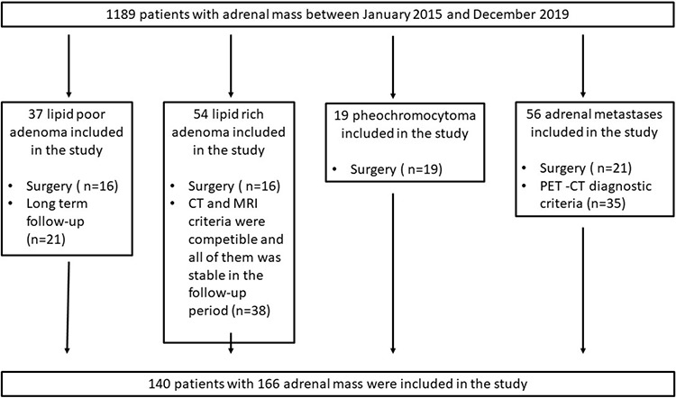
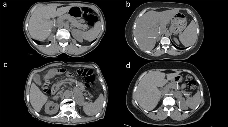
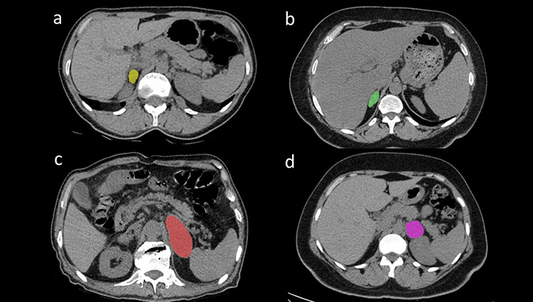
Methods: In this single-center retrospective study, 166 adrenal lesions in 140 patients (64 women, 76 men; mean age 56.58 ± 13.65 years) were evaluated between January 2015 and December 2019. The lesions consisted of 54 lipid-rich adrenal adenomas, 37 lipid-poor adrenal adenomas (LPAs), 56 adrenal metastases (ADM), and 19 adrenal pheochromocytomas (APhs). Each adrenal lesion was segmented by manually contouring the borders of the lesion on unenhanced CT images. A texture analysis of the CT images was performed using Local Image Feature Extraction software. First-order and second-order texture parameters were assessed, and 45 features were extracted from each lesion. One-Way analysis of variance with Bonferroni correction and the Mann-Whitney U test was performed to determine the relationships between the texture features and adrenal lesions. Receiver operating characteristic curves were performed for lesion discrimination based on the texture features. Logistic regression analysis was used to generate logistic models, including only the texture parameters with a high-class separation capacity (i.e., P < 0.050). SPSS software was used for all statistical analyses.
Results: First-order and second-order texture parameters were identified as significant factors capable of differentiating among the four lesion types (P < 0.050). The logistic models were evaluated to ascertain the relationships between LPA and ADM, LPA and APh, and ADM and APh. The sensitivity, specificity, positive predictive value (PPV), negative predictive value (NPV), and accuracy of the first model (LPA vs. ADM) were 85.7%, 70.3%, 81.3%, 76.4%, and 79.5%, respectively. The sensitivity, specificity, PPV, NPV, and accuracy of the second model (LPA vs. APh) were all 100%. The sensitivity, specificity, PPV, NPV, and accuracy of the third model (ADM vs. APh) were 87.5%, 82%, 36.8%, 98.2%, and 82.7%, respectively.
Conclusion: Texture features may help in the characterization of adrenal lesions on unenhanced CT images.
Keywords: Adrenal adenoma; adrenal glands; adrenal mass; computed tomography; texture analysis.
Conflict of interest statement
The authors declared no conflicts of interest.
Figures
References
-
- Shi B, Zhang GM, Xu M, Jin ZY, Sun H. Distinguishing metastases from benign adrenal masses: what can CT texture analysis do? Acta Radiol. 2019;60(11):1553–1561. - PubMed
-
- Song JH, Chaudhry FS, Mayo-Smith WW. The incidental indeterminate adrenal mass on CT (> 10 H) in patients without cancer: is further imaging necessary? Follow-up of 321 consecutive indeterminate adrenal masses. AJR Am J Roentgenol. 2007;189(5):1119–1123. - PubMed
-
- Schieda N, Siegelman ES. Update on CT and MRI of Adrenal Nodules. AJR Am J Roentgenol. 2017;208(6):1206–1217. - PubMed
-
- Bharwani N, Rockall AG, Sahdev A, et al. Adrenocortical carcinoma: the range of appearances on CT and MRI. AJR Am J Roentgenol. 2011;196(6):W706–W714. - PubMed
MeSH terms
Substances
LinkOut - more resources
Full Text Sources
Medical
Miscellaneous
