. 2023 Apr 21;18(4):756-771.
doi: 10.1021/acschembio.2c00720. Epub 2023 Mar 29.

Synthetic ERRα/β/γ Agonist Induces an ERRα-Dependent Acute Aerobic Exercise Response and Enhances Exercise Capacity

Affiliations

Synthetic ERRα/β/γ Agonist Induces an ERRα-Dependent Acute Aerobic Exercise Response and Enhances Exercise Capacity

Cyrielle Billon et al. ACS Chem Biol. .

Abstract

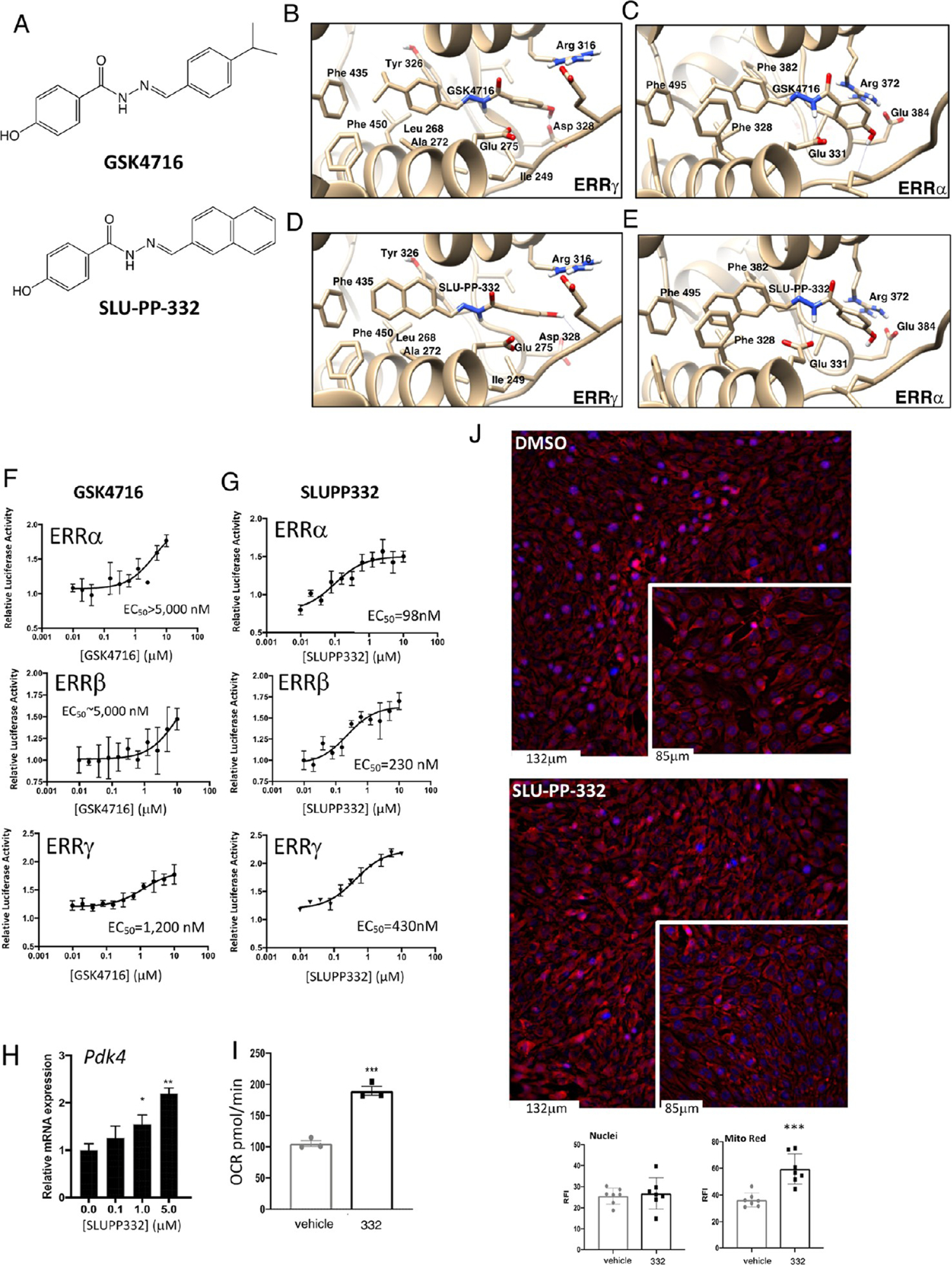
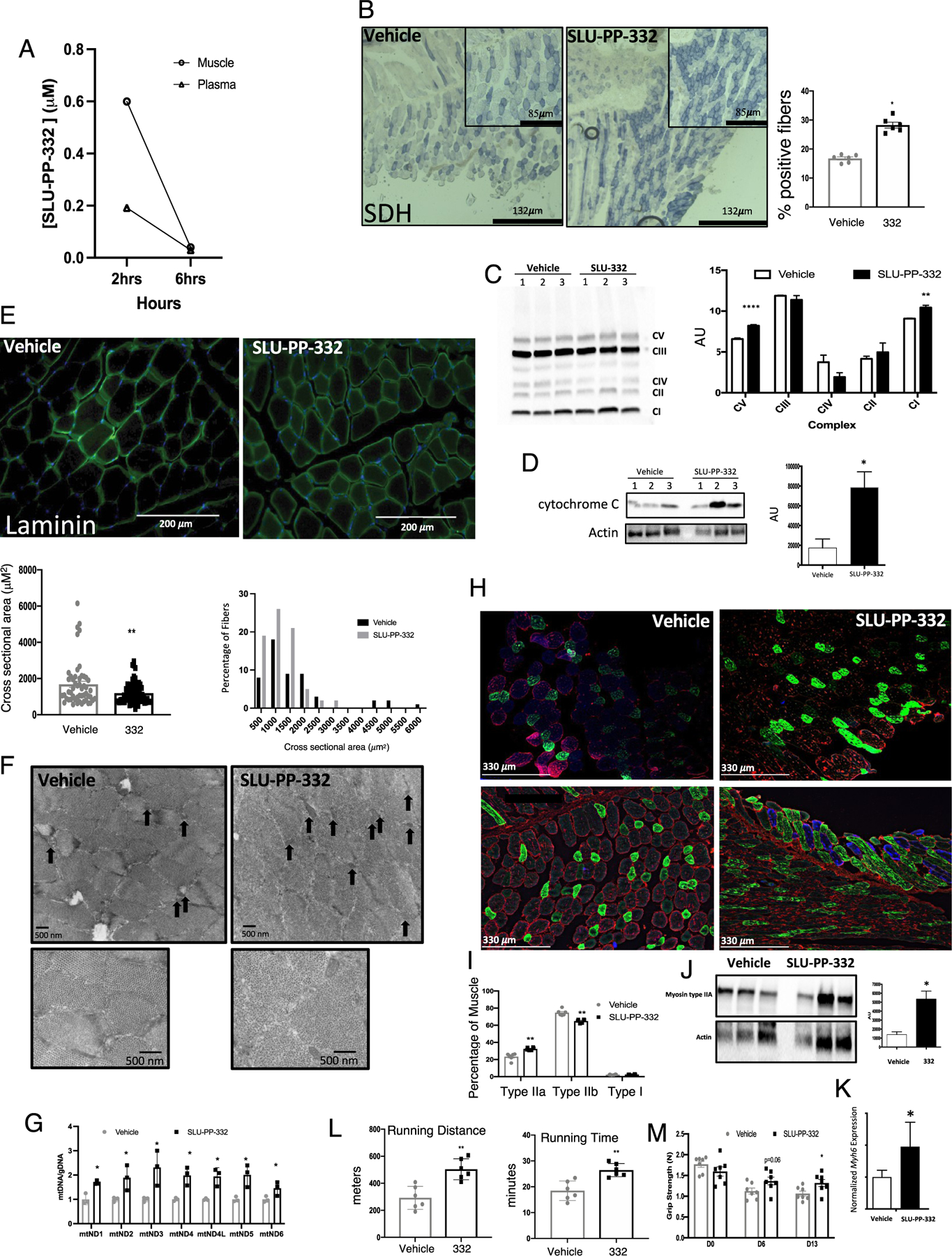
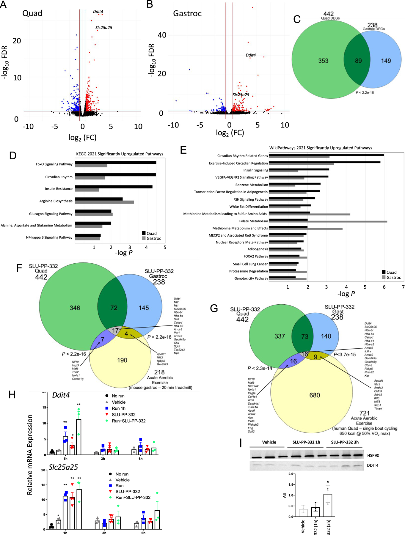
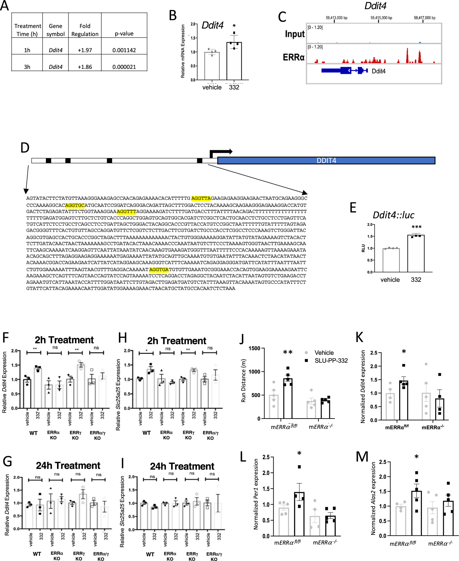
Repetitive physical exercise induces physiological adaptations in skeletal muscle that improves exercise performance and is effective for the prevention and treatment of several diseases. Genetic evidence indicates that the orphan nuclear receptors estrogen receptor-related receptors (ERRs) play an important role in skeletal muscle exercise capacity. Three ERR subtypes exist (ERRα, β, and γ), and although ERRβ/γ agonists have been designed, there have been significant difficulties in designing compounds with ERRα agonist activity. Additionally, there are limited synthetic agonists that can be used to target ERRs in vivo. Here, we report the identification of a synthetic ERR pan agonist, SLU-PP-332, that targets all three ERRs but has the highest potency for ERRα. Additionally, SLU-PP-332 has sufficient pharmacokinetic properties to be used as an in vivo chemical tool. SLU-PP-332 increases mitochondrial function and cellular respiration in a skeletal muscle cell line. When administered to mice, SLU-PP-332 increased the type IIa oxidative skeletal muscle fibers and enhanced exercise endurance. We also observed that SLU-PP-332 induced an ERRα-specific acute aerobic exercise genetic program, and the ERRα activation was critical for enhancing exercise endurance in mice. These data indicate the feasibility of targeting ERRα for the development of compounds that act as exercise mimetics that may be effective in the treatment of numerous metabolic disorders and to improve muscle function in the aging.


Conflict of interest statement

The authors declare the following competing financial interest(s): T.P.B., B.E., and J.K.W. are stockholders in Myonid Therapeutics, Inc., which focuses on ERR based therapeutics.

Figures

Figure 1.
Figure 1.
SLU-PP-332 is a Pan ERRα/β/γ agonist with significant ERRα activity. (A) Chemical structures of GSK4716 (top) and SLU-PP-332 (bottom). (B) Schematic illustrating the X-ray co-crystal structure of GSK4716 bound to the ERRγ LBD (PDB ID: 2GPP). Models of ERRα bound to GSK4716 (C), ERRγ LBD bound to GSK4716 (D), and ERRα bound to SLU-PP-332 (E). Results of ERRα, ERRβ, or ERRγ cotransfection assays and full-length ERRs in HEK293 cells illustrating the activity of GSK4716 (F) and SLU-PP-332 (G). (H) Effects of SLU-PP-332 dose–response treatment (24 h) on pyruvate dehydrogenase kinase 4 (Pdk4), in C2C12 cells, n = 3. (I) Maximal mitochondrial respiration values analyzed by Seahorse of C2C12 cells treated with dimethyl sulfoxide (DMSO) (gray bar) or SLU-PP-332 (black bar), n = 3. (J) MitoTracker Red staining of C2C12 cells under proliferative conditions treated with DMSO or SLU-PP-332 (10 μM) for 24 h. Bar graph represents nuclei intensity (left) and MitoTracker Red staining (right). p < 0.05, **p < 0.01, and ***p < 0.05.
Figure 2.
Figure 2.
SLU-PP-332 increases oxidative fibers in skeletal muscle and improves exercise endurance. (A) Pharmacokinetic analysis of SLU-PP-332 displaying muscle and plasma levels of the compound at 2 and 6 h post 30 mg/kg, i.p. in 10/10/80 DMSO, Tween, phosphate-buffered saline (PBS). n = 3. (B) Immunochemical analysis of succinate dehydrogenase (SDH) from quadriceps of mice administered vehicle (white bars, n = 8) or SLU-PP-332 50 mg/kg, b.i.d (black bar, n = 8). The bar graph represents the quantification of SDH-positive muscle fibers. (C) OXPHOS complex blot from quadriceps of mice dosed with vehicle or SLU-PP-332 50 mg/kg, b.i.d (black bar, n = 7). Protein normalization was performed using the Stain-Free Western Workflow suite from Bio-Rad. Relative amount of total protein in each lane on the blot was calculated and used for quantitation normalization. We then identified a band for each OXPHOS complex and then normalized their intensity level to the total protein intensity for each lane. Then, the average and standard deviation were calculated for each group (vehicle and SLU-332). (D) Cytochrome c protein levels from quadriceps from mice dosed with vehicle (white bars, n = 7) or SLU-PP-332 50 mg/kg, b.i.d (black bar, n = 7). The bar graph represents the quantification of expression. Immunochemical analysis of laminin (E), electron microscopy of quadriceps (F) (black arrows illustrate identified mitochondria). (G) Analysis of mitochondrial DNA levels (relative to nuclear DNA) from quadriceps. Immunochemical analysis of muscle fiber types. (H) Stained sections (n = 6 per group) from quadriceps of mice administered vehicle (white bars, n = 8) or SLU-PP-332 50 mg/kg, b.i.d (black bar, n = 8). Myosin IIa is green, myosin IIb is red, and myosin I is blue. The bar graph represents the quantification of fiber cross-sectional area (lower panel) and percentage of fiber types (I). Myosin heavy chain protein (J) and gene (K) expression from quadriceps of mice treated with vehicle or SLU-PP-332 50 mg/kg, b.i.d (n = 6 for gene expression and n = 3 for protein). For (J), myosin IIA protein expression was normalized to actin after imaging and dividing the myosin IIA protein signal to the respective actin signal using ImageJ software. The bar graph represents the quantification of expression. (L) Running distance (left panel) and running time (right panel) of mice treated with an acute dose of vehicle (gray bar) or SLU-PP-332 (50 mg/kg, black bar) for 1 h before running (n = 6 per group). (M) Grip strength test from mice treated with vehicle (gray bar) or SLU-PP-332 (50 mg/kg, black bar) before dosing (D0), after 6 days of dosing (D6), or after 13 days (D13) (n = 8). p < 0.05, **p < 0.01, ***p < 0.001, and ****p < 0.0001.
Figure 3.
Figure 3.
SLU-PP-332 induces acute aerobic exercise genetic program in skeletal muscles. Results from RNA-seq studies from the gastrocnemius or quadricep muscles of mice treated with vehicle or SLU-PP-332. (A) Volcano plot illustrating differentially expressed genes (DEGs) (FDR > 0.05, | FC| > 1.5) from the quadriceps of SLU-PP-332-treated mice. Muscles were collected 3 h post final dose of a 10-day dosing regimen 50 mg/kg b.i.d. (B) Volcano plot illustrating differentially expressed genes (DEGs) (FDR > 0.05, |FC| > 1.5) from the gastrocnemius muscle of SLU-PP-332-treated mice. Muscles were collected 3 h post final dose of a 10-day dosing regimen 50 mg/kg b.i.d. (C) Venn diagram representing the overlap of DEGs from the two muscle types shown in panels (A) and (B). (D, E) Pathway analysis of DEGs in quadricep and gastrocnemius muscles after treatment with SLU-PP-332 in mice. The significantly (p < 0.05) regulated pathways shared in both muscle types are listed for KEGG pathways (D) and Wikipathways (E). (F) Venn diagram illustrating the overlap of SLU-PP-332 DEGs and DEGs identified in the gastrocnemius muscle of mice subjected to acute aerobic exercise (see text for details). Fisher’s exact test was performed to determine significance. (G) Venn diagram illustrating the overlap of SLU-PP-332 DEGs and DEGs identified in the quadricep muscle of lean humans subjected to acute aerobic exercise (see text for details). Fisher’s exact test was performed to determine significance. (H) Ddit4 (upper panel) and Slc25c25 expression (lower panel) from quadriceps from mice treated with vehicle (gray triangle), SLU-PP-332 (50 mg/kg, i.p., red triangle), SLU-PP-332 in combination of 45 min of running (green circle), or no treatment (black circle). Mice were euthanized as indicated 1, 3, or 6 h after treatment (n = 6 per group). (I) DDIT4 protein expression from quadriceps from mice treated with SLU-PP-332 (50 mg/kg, i.p.). Mice were euthanized as indicated 1 or 3 h after treatment (n = 3 per group). The bar graph represents the quantification of expression. p < 0.05, **p < 0.01, ***p < 0.001, and ****p < 0.0001.
Figure 4.
Figure 4.
SLU-PP-332 induces Ddit4 expression and enhances exercise endurance in an ERRα-dependent manner. (A) Effects of SLU-PP-332 treatment (10 μM) for either 1 or 3 h on Ddit4 expression in C2C12 cells detected using the Qiagen RT PCR array. (B) Effects of SLU-PP-332 (10 μM) treatment of C2C12 for 3 h gene on Ddit4 expression (n = 3). (C) ERRα binding locations near and within the Ddit4 gene identified by ChIP-seq in C2C12 cells. (D) Schematic representation of luciferase reporter containing the putative ERRα binding site from Ddit4 identified in (C). (E) Cotransfection assay in HEK293T cells with full-length ERRα (including SLU-PP-332 (10 μM)). Ddit4 and Slc25a25 expression are transiently induced in primary myocytes by SLU-PP-332 in an ERRα-dependent manner. Ddit4 expression in primary ERR WT, ERRα KO, ERRγ KO, and ERRα/γ KO myoblasts treated with DMSO or SLU-PP-332 (1 μM) for 2 h (F) or 24 h (G). Slc25a25 expression in primary ERR WT, ERRα KO, ERRγ KO, and ERRα/γ KO myoblasts treated with DMSO or SLU-PP-332 (1 μM) for 2 h (H) or 24 h (I). (J) Running endurance of mERRαfl/fl vs mERRα−/− mice dosed with vehicle or SLU-PP-332. (K) Expression of Ddit4 mRNA (quadricep) mice shown in panel (J) measured by QPCR (L) Expression of Per1 mRNA (quadricep) mice shown in panel (J) measured by QPCR (M) Expression of Alas2 mRNA (quadricep) mice shown in panel (J) measured by QPCR. p < 0.05, **p < 0.01, ***p < 0.001, and ****p < 0.0001.

References

    1. Booth FW; Roberts CK; Laye MJ Lack of exercise is a major cause of chronic diseases. Compr. Physiol 2011, 2, 1143–1211. - PMC - PubMed
    1. O’Gorman DJ; Karlsson HK; McQuaid S; Yousif O; Rahman Y; Gasparro D; Glund S; Chibalin AV; Zierath JR; Nolan JJ Exercise training increases insulin-stimulated glucose disposal and GLUT4 (SLC2A4) protein content in patients with type 2 diabetes. Diabetologia 2006, 49, 2983–2992. - PubMed
    1. Diabetes Prevention Program Research Group. Reduction in the incidence of type 2 diabetes with lifestyle intervention or metformin. N. Engl. J. Med 2002, 346, 393–403. - PMC - PubMed
    1. Koopman R; Manders RJ; Zorenc AH; Hul GB; Kuipers H; Keizer HA; van Loon LJ A single session of resistance exercise enhances insulin sensitivity for at least 24 h in healthy men. Eur. J. Appl. Physiol 2005, 94, 180–187. - PubMed
    1. Borsheim E; Bahr R Effect of exercise intensity, duration and mode on post-exercise oxygen consumption. Sports Med 2003, 33, 1037–1060. - PubMed

Publication types

MeSH terms

LinkOut - more resources