Case Reports
. 2023 Mar 1;50(2):e227922.
doi: 10.14503/THIJ-22-7922.

Large Left Ventricular Pseudoaneurysm Presenting as an Embolic Stroke After a "Silent" Myocardial Infarction

Affiliations
Case Reports

Large Left Ventricular Pseudoaneurysm Presenting as an Embolic Stroke After a "Silent" Myocardial Infarction

Cameron Incognito et al. Tex Heart Inst J. .

Abstract

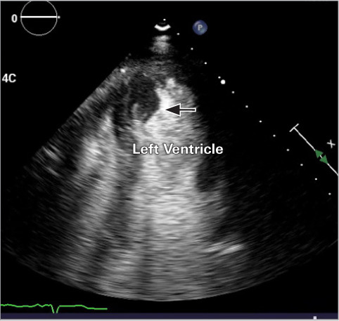
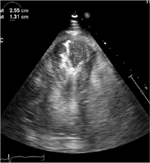
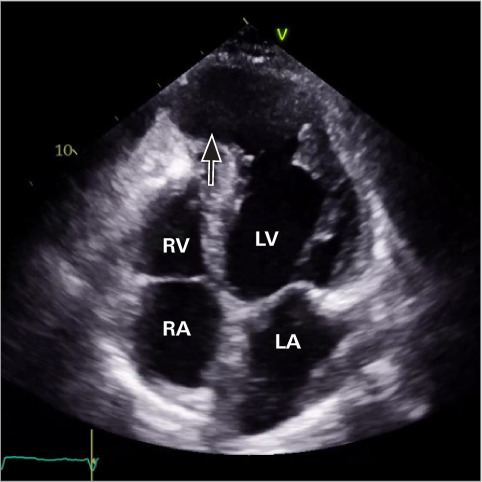
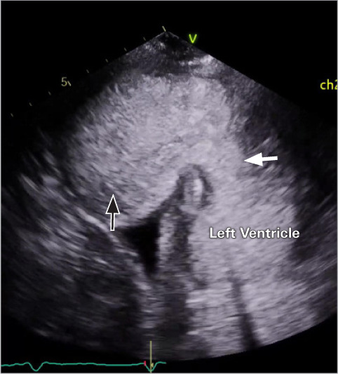
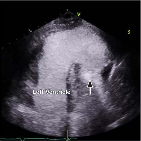
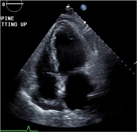
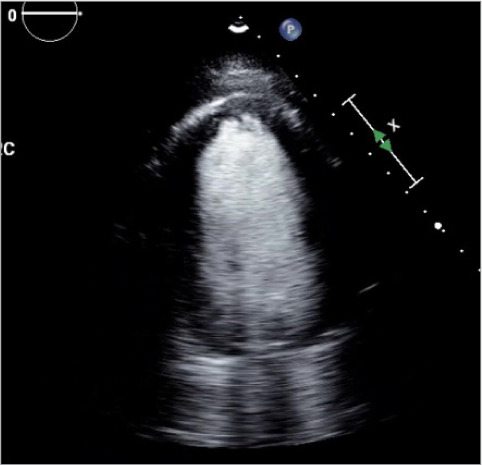
A 72-year-old woman with no history of coronary artery disease presented with an acute left middle cerebral artery stroke and was found to have a large left ventricular pseudoaneurysm measuring 8.7 × 7.6 cm and 2 large left ventricular thrombi, the source of her systemic embolization. Despite initial medical management, she developed refractory New York Heart Association functional class III heart failure, uncontrolled atrial fibrillation, and further enlargement of her pseudoaneurysm to 5.5 × 10.6 × 9.2 cm. She underwent urgent aneurysmectomy. Left ventricular pseudoaneurysms are rare and most commonly occur following an acute myocardial infarction when a ventricular free-wall rupture is contained by pericardium or thrombi. Historically, left ventricular angiography displaying a lack of an overlying coronary artery was the gold standard for diagnosis. Now, noninvasive imaging such as computed tomography, magnetic resonance imaging, and echocardiogram with ultrasound-enhancing agent, are reliable diagnostic tools. They can distinguish a pseudoaneurysm from a true left ventricular aneurysm using characteristic findings such as a narrow aneurysm neck, bidirectional doppler flow between the pseudoaneurysm and the left ventricle, and abrupt changes in the cardiac wall structures. Progressive dilation, wall thinning, and dyskinesis can result in refractory heart failure, arrhythmias, and thrombi formation from venous stasis. Pseudoaneurysms have a 30% to 45% risk of rupture and can be treated with left ventricular aneurysmectomy.

Keywords: Pseudoaneurysm; embolic stroke; myocardial infarction.


Conflict of interest statement

Conflict of Interest Disclosures: None.

Figures

Fig. 1
Fig. 1
Initial apical 2-chamber transthoracic echocardiogram with an ultrasound-enhancing agent shows a thrombus measuring 2.7 × 1.3 cm (arrow) within the apex of the left ventricle. Not visualized is an additional 1.25 × 0.85 cm thrombus and the pseudoaneurysm measuring 8.7 × 7.6 cm. Supplemental motion image is available for Figure 1.
Fig. 2
Fig. 2
Initial apical 2-chamber transthoracic echocardio-gram with an ultrasound-enhancing agent shows both thrombi, measuring 1.25 × 0.85 cm (left) and 2.7 × 1.3 cm (right), within the apex of the left ventricle.
Fig. 3
Fig. 3
Repeat apical 4-chamber transthoracic echocardio-gram without UEA shows a large apical pseudoaneurysm (arrow) that measured 10.6 × 9.2 cm, 5 months after the index admission. The 2 prior thrombi had resolved but were also not visualized using UEA (thus not shown). Supplemental motion image is available for Figure 3. LA, left atrium; LV, left ventricle; RA, right atrium; RV, right ventricle; UEA, ultrasound-enhancing agent.
Fig. 4
Fig. 4
Repeat apical 2-chamber transthoracic echocardio-gram with an ultrasound-enhancing agent shows a large apical pseudoaneurysm (black arrow; 10.6 × 9.2 cm) with a narrow neck (white arrow), 5 months after the index admission. Supplemental motion image is available for Figure 4.
Fig. 5
Fig. 5
Repeat apical long-axis transthoracic echocardio-gram with an ultrasound-enhancing agent shows a large apical pseudoaneurysm (arrow; 10.6 × 9.2 cm), 5 months after the index admission. Supplemental motion image is available for Figure 5.
Fig. 6
Fig. 6
Postoperative day 3 apical 4-chamber transthoracic echocardiogram without an ultrasound-enhancing agent following aneurysmectomy, with an estimated left ventricular ejection fraction of 33% and an left ventricular end-diastolic diameter of 6.7 cm. Supplemental motion image is available for Figure 6.
Fig. 7
Fig. 7
Postoperative day 3 apical 2-chamber transthoracic echocardiogram with ultrasound-enhancing agent following aneurysmectomy, with estimated left ventricular ejection fraction of 33% and an left ventricular end-diastolic diameter of 6.7 cm. Supplemental motion image is available for Figure 7.

References

    1. Brown SL, Gropler RJ, Harris KM. Distinguishing left ventricular aneurysm from pseudoaneurysm. A review of the literature. Chest . 1997;111(5):1403–1409. doi: 10.1378/chest.111.5.1403. - DOI - PubMed
    1. Ruzza A, Czer LS, Arabia F et al. Left ventricular reconstruction for postinfarction left ventricular aneurysm: review of surgical techniques. Tex Heart Inst J . 2017;44(5):326–335. doi: 10.14503/THIJ-16-6068. - DOI - PMC - PubMed
    1. Frances C, Romero A, Grady D. Left ventricular pseudoaneurysm. J Am Coll Cardiol . 1998;32(3):557–561. doi: 10.1016/s0735-1097(98)00290-3. - DOI - PubMed
    1. Alapati L, Chitwood WR, Cahill J, Mehra S, Movahed A. Left ventricular pseudoaneurysm: a case report and review of the literature. World J Clin Cases . 2014;2(4):90–93. doi: 10.12998/wjcc.v2.i4.90. - DOI - PMC - PubMed
    1. March KL, Sawada SG, Tarver RD, Kesler KA, Armstrong WF. Current concepts of left ventricular pseudoaneurysm: pathophysiology, therapy, and diagnostic imaging methods. Clin Cardiol . 1989;12(9):531–540. doi: 10.1002/clc.4960120911. - DOI - PubMed

Publication types