. 2023 Mar 29;9(13):eade1792.
doi: 10.1126/sciadv.ade1792. Epub 2023 Mar 29.

mRNA translational specialization by RBPMS presets the competence for cardiac commitment in hESCs

Affiliations

mRNA translational specialization by RBPMS presets the competence for cardiac commitment in hESCs

Deniz Bartsch et al. Sci Adv. .

Abstract

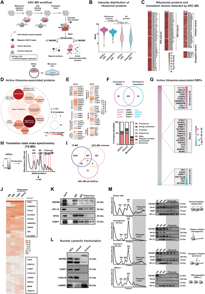
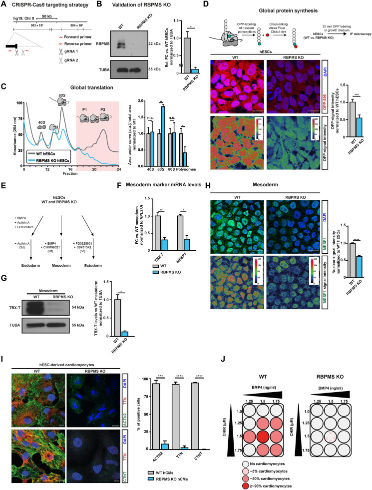
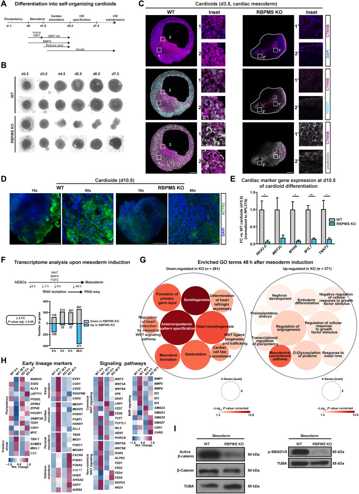
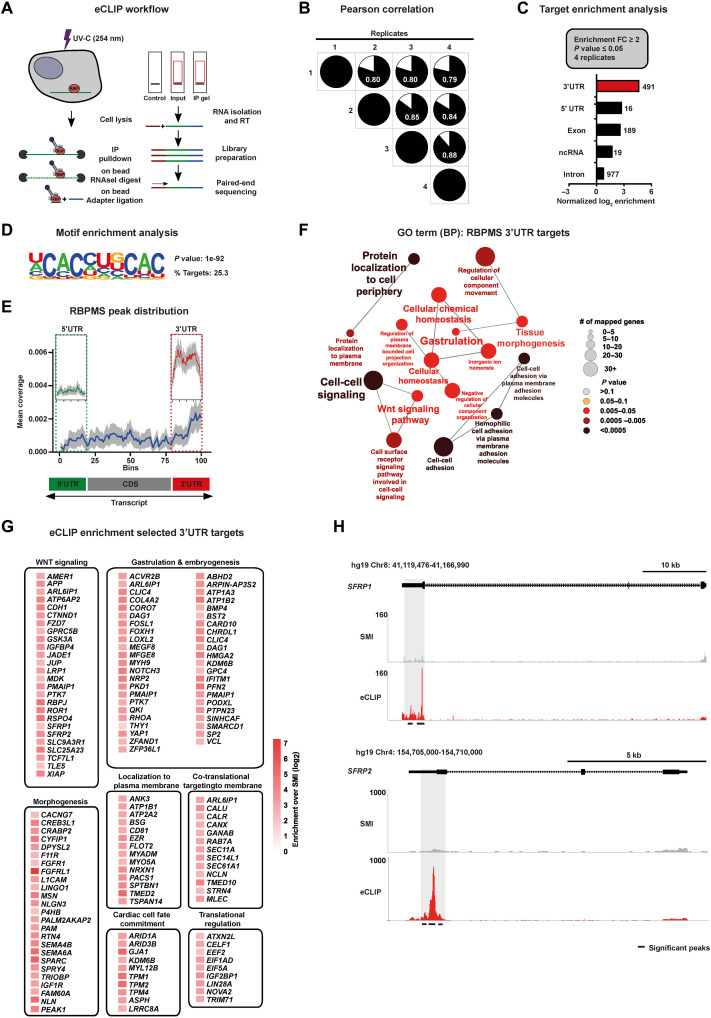
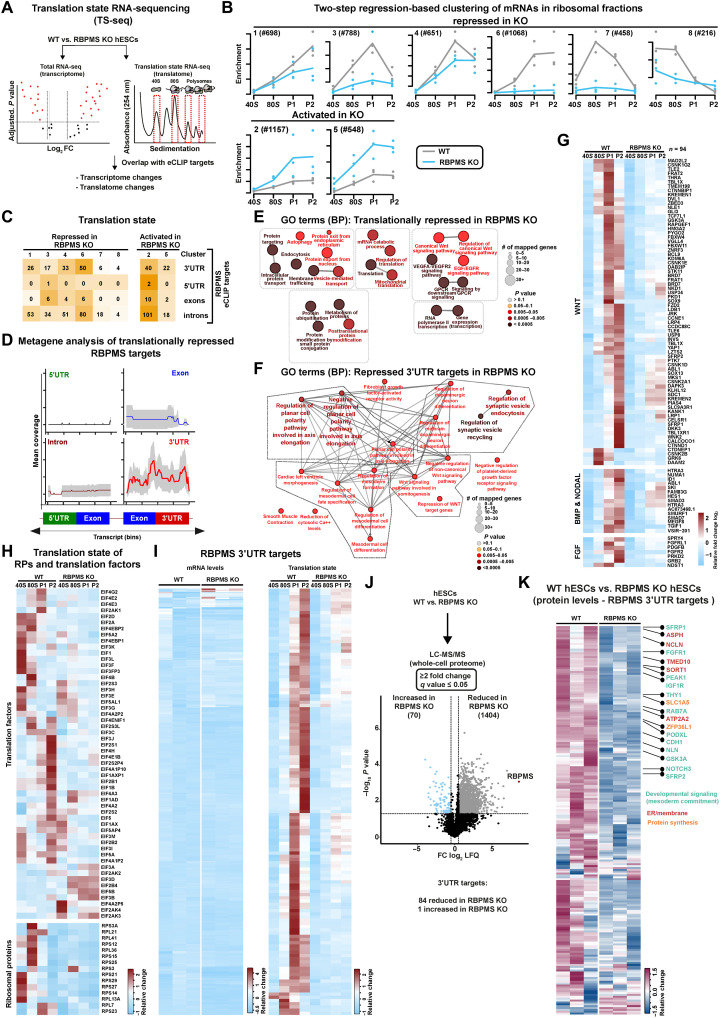
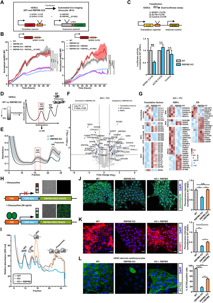
The blueprints of developing organs are preset at the early stages of embryogenesis. Transcriptional and epigenetic mechanisms are proposed to preset developmental trajectories. However, we reveal that the competence for the future cardiac fate of human embryonic stem cells (hESCs) is preset in pluripotency by a specialized mRNA translation circuit controlled by RBPMS. RBPMS is recruited to active ribosomes in hESCs to control the translation of essential factors needed for cardiac commitment program, including Wingless/Integrated (WNT) signaling. Consequently, RBPMS loss specifically and severely impedes cardiac mesoderm specification, leading to patterning and morphogenetic defects in human cardiac organoids. Mechanistically, RBPMS specializes mRNA translation, selectively via 3'UTR binding and globally by promoting translation initiation. Accordingly, RBPMS loss causes translation initiation defects highlighted by aberrant retention of the EIF3 complex and depletion of EIF5A from mRNAs, thereby abrogating ribosome recruitment. We demonstrate how future fate trajectories are programmed during embryogenesis by specialized mRNA translation.


Figures

Fig. 1.
Fig. 1.. ARC-MS identifies proteins recruited to translationally active ribosomes during mesoderm commitment.
(A) Schematic of ARC-MS workflow. ARC-MS was performed in hESCs (d0) and hESC-derived mesoderm progenitors (d2). (B) Violin plots depicting intensity-based absolute quantification (IBAQ) values of ribosomal proteins and all identified proteins from ARC-MS data from hESCs versus mesoderm progenitors. (C) Heatmap showing the enrichment of ribosomal proteins and translation factors (EIFs and EEFs) detected by ARC-MS in hESCs and mesoderm progenitors. (D) GO-based functional enrichment analysis for proteins (excluding ribosomal proteins and translation factors) reliably identified by ARC-MS. (E) Heatmap depicting log2 LFQ of significantly enriched representative proteins recruited on active ribosomes from major GO term categories identified by ARC-MS. (F) Venn diagram summarizing the distribution of proteins on active ribosomes in hESCs and mesoderm progenitors. Identified proteins, categorized based on molecular function, are depicted as a percentage of the total in the bar graphs below. (G) Heatmap displaying the enrichment of RBPs identified by ARC-MS between d0 and d2. (H) Schematic outline of the underlying strategy used for TS-MS. (I) Overlap of proteins enriched at d0 and d2 ARC-MS with proteins detected via TS-MS in hESCs. (J) Distribution of proteins recruited on active ribosomes selectively in hESCs identified by ARC-MS that are overlapping with TS-MS on indicated ribosomal fraction. (K) Confirmation of RBPMS enrichment in ribosomal fractions by polysome profiling followed by immunoblotting. RPL7A, RPS6, and G3BP1 = controls. (L) RBPMS is predominantly a cytosolic protein in hESCs, evaluated by Western blot analysis upon nuclear/cytosolic fractionation, G3BP1, and TUBA cytosolic control, LAMINB1 nuclear control. (M) Residence of RBPMS on ribosomal complexes confirmed by polysome profiling upon treatment with the indicated translation inhibitors. Error bars represent ±SEM; P values are calculated using Student’s t test; biological replicates n = 3.
Fig. 2.
Fig. 2.. RBPMS loss causes global translation inhibition in hESCs without affecting self-renewal and selectively impedes cardiac mesoderm specification.
(A) Schematic representation of RBPMS locus in humans and the CRISPR-Cas9–based targeting strategy used to generate homozygous RBPMS-KO (B) confirmed via immunoblot. (C) Loss of RBPMS impedes translation in hESCs, indicated by representative polysome profiles of RBPMS-KO hESCs with respect to isogenic WT along with quantification of the area under the indicated ribosomal fractions on the right. (D) De novo protein synthesis is inhibited upon RBPMS loss, evaluated by measuring puromycin incorporation on nascent polypeptides in RBPMS-KO compared to WT by measuring uptake of OPP (quantifications on the right). (E) Schematic of lineage differentiation approaches used to determine the competence of RBPMS-KO hESCs to undergo germline commitment. (F) Mesoderm commitment is severely impaired upon loss of RBPMS as indicated by reverse transcription-quantitative polymerase chain reaction (RT-qPCR) for TBX-T (mesoderm) and MESP1 (cardiac mesoderm), as well as (G) Western blot for TBX-T (quantification on the right). (H) Representative images of MESP1 staining upon mesoderm induction of RBPMS-KO hESCs compared to WT (quantification on the right). (I) Immunofluorescence images for cardiac-specific ACTN2 and TTN. The bar graph shows normalized expression levels of indicated cardiomyocyte markers. (J) Schematic summarizing the cardiac differentiation efficiency along the cardiac corridor for WT and RBPMS-KO, indicating the inability of hESCs to terminally differentiate to cardiomyocytes upon RBPMS loss. Error bars represent ±SEM; P values calculated using Student’s t test (*P ≤ 0.05, **P ≤ 0.01, ***P ≤ 0.001, and ****P ≤ 0.0001; n = 3).
Fig. 3.
Fig. 3.. RBPMS is essential for cardiac mesoderm patterning and morphogenesis in human cardioids.
(A) Schematic of cardioid generation method. (B) Loss of RBPMS impairs cardioid formation at early stages of cardiogenesis as indicated by bright-field images taken at indicated days during cardiac induction. (C) Whole-organoid confocal imaging for HAND1, CTNNB, and phalloidin at cardiac mesoderm stage cardioids (d3.5) derived from RBPMS-KO and WT hESCs. Stitched images of the whole organoid acquired by 63× objective are shown. (D) Whole-organoid imaging for cardiomyocyte-specific ACTN2 in WT and RBPMS-KO cardioids (d10.5). (E) RT-qPCR analysis for mRNA levels of cardiac-specific transcription factors and sarcomeric proteins in the indicated samples, performed on cardioids at d10.5. (F) Systematic identification of differentially expressed genes at indicated early and late time points during mesoderm induction upon RBPMS loss (FC ± 2, P ≤ 0.05, n = 3) evaluated by RNA-seq, represented as a bar graph. (G) GO-based analysis of genes down-regulated and up-regulated at 48 hours after mesoderm induction indicates defects in mesoderm and cardiac mesoderm cell fate specification and WNT signaling in RBPMS-KO cells w.r.t WT. (H) Heatmaps showing the expression of cell fate markers (first two panels) and the components of WNT and BMP signaling in pluripotency and upon mesoderm induction of WT and RBPMS-KO. (I) Absence of RBPMS impairs WNT signaling activity, indicated by active β-catenin levels, and BMP signaling, indicated by pSMAD1/5, upon cardiac mesoderm induction of WT and RBPMS-KO. Error bars represent ±SEM; P values calculated using Student’s t test (*P ≤ 0.05, **P ≤ 0.01, ***P ≤ 0.001, and ****P ≤ 0.0001; n = 3).
Fig. 4.
Fig. 4.. mRNAs encoding cardiac mesoderm regulators are targeted by RBPMS via 3′UTR binding.
(A) Schematic of the eCLIP-seq approach used to faithfully generate a transcriptome-wide direct binding map for RBPMS at single-nucleotide resolution. (B) Biological quadruplicates of RBPMS eCLIP-seq show at least 80% overlap. Pie charts show the correlation of statistically significant uniquely mapped reads for each replicate over SMInput. (C) RBPMS reliably binds predominantly the 3′UTR of transcripts, demonstrated here by the distribution of the significantly enriched eCLIP peaks against the paired SMInput (FC ≥ 2; P ≤ 0.05 in all four replicates). (D) Top sequence motif significantly bound by RBPMS. (E) Metagene plot visualizing the RBPMS peak distribution over SMInput illustrating prominent 3′UTR binding. (F) 3′UTR targets of RBPMS regulate molecular processes central to mesoderm/cardiac commitment, including WNT signal transduction, depicted by significantly enriched GO terms. (G) A curated set of RBPMS 3′UTR targets grouped based on their proven role in the indicated cellular, developmental, and functional process, depicted as a heatmap of fold enrichment over SMInput. (H) Representative read density tracks show read density for RBPMS across the gene body of SFRP1 and SFRP2, a representative target.
Fig. 5.
Fig. 5.. RBPMS controls mRNA translation of factors essential to initiate cardiac mesoderm commitment.
(A) Schematic of the TS-seq strategy used to evaluate the translational status of RBPMS-KO compared to WT (n = 3). (B) Global impact of the loss of RBPMS on ribosome occupancy in hESCs, revealed by two-step regression analysis of the mRNAs enriching on indicated ribosomal fractions derived from TS-seq. (C) Translation state of mRNAs bound by RBPMS in RBPMS-KO compared to WT, grouped based on RBPMS binding coordinates, in the indicated translationally affected clusters identified by TS-seq. (D) Metagene plot revealing RBPMS 3′UTR binding bias for translationally repressed RBPMS targets. (E) Functional analysis of all translationally repressed mRNAs and (F) translationally repressed 3′UTR targets in RBPMS-KO versus WT hESCs illustrated as a significantly enriched, curated list of GO terms. (G) Loss of RBPMS severely inhibits translation of the components of vital mesoderm specifying signal transduction networks (WNT, BMP, NODAL, and FGF signaling), as well as (H) translation factors and ribosomal proteins. (I) mRNAs bound by RBPMS at the 3′UTR are depleted from ribosomes in RBPMS-KO without affecting the transcript levels. The heatmap on the left depicts mRNA levels of RBPMS 3′UTR targets, while the heatmap on the right depicts their ribosome occupancy. (J) Changes in total proteome between WT hESCs and RBPMS-KO hESCs depicted as a volcano plot derived from whole-cell proteomics analysis. (K) Heatmap depicting protein levels (as log2 LFQ values) of RBPMS 3′UTR targets in WT and RBPMS-KO hESCs.
Fig. 6.
Fig. 6.. RBPMS specializes mRNA translation in pluripotency, selectively via 3′UTR binding and globally by controlling translation initiation and ribosome recruitment.
(A) Schematic of the reporter system and the experimental workflow used to investigate the 3′UTR binding motif–dependent regulation of translation by RBPMS in hESCs. (B) RBPMS activates translation of reporter mRNA carrying RBPMS binding motifs in the 3′UTR, evaluated by time-lapse microscopy. (C) The presence of the RBPMS binding motif is required for 3′ binding–dependent translation activation by RBPMS evaluated using indicated luciferase-based bicistronic reporters. (D) Schematic outlining the translation complex profiling–based isolation of 40S and preinitiation complex (PIC), followed by proteomics analysis in WT and RBPMS-KO hESCs. (E) Translation complex profiling traces of WT and RBPMS-KO hESCs (shades represent SEM). 40S + PIC fractions were subjected to LC-MS/MS. (F) Proteins significantly changing in the 40S + PIC fraction between WT and RBPMS-KO hESCs represented as volcano plot. Dashed lines indicate significance thresholds (−log10 P ≥ 1.3 and log2 FC ± 2) (selected translation factors, ER proteins, and RBPs are highlighted by orange, green, and blue dots, respectively). (G) Heatmap depicting differentially enriched translation factors, RBPs, and translation-associated ER proteins in the 40S + PIC fraction between WT and RBPMS-KO hESCs (significantly changing proteins are highlighted in bold). (H) Illustration of the PiggyBac-based strategy used to reexpress RBPMS in RBPMS-KO. Representative microscopy images in the inlets before and after induction. Timely reconstitution of RBPMS in RBPMS-KO hESCs rescues (I) translation defects, (J) protein synthesis defects, (K) translation defect of representative 3′UTR target of RBPMS, SFRP1, and (L) cardiac differentiation defect. Quantification of the microscopy images on the right side as bar graphs. Error bars represent ±SEM; P values calculated using Student’s t test (*P ≤ 0.05, **P ≤ 0.01, ***P ≤ 0.001, and ****P ≤ 0.0001; n = 3).

References

    1. J. Newport, M. Kirschner, A major developmental transition in early Xenopus embryos: II. Control of the onset of transcription. Cell 30, 687–696 (1982). - PubMed
    1. J. Newport, M. Kirschner, A major developmental transition in early Xenopus embryos: I. characterization and timing of cellular changes at the midblastula stage. Cell 30, 675–686 (1982). - PubMed
    1. H. Spemann, H. Mangold, Induction of embryonic primordia by implantation of organizers from a different species. 1923. Int. J. Dev. Biol. 45, 13–38 (2001). - PubMed
    1. L. Xiang, Y. Yin, Y. Zheng, Y. Ma, Y. Li, Z. Zhao, J. Guo, Z. Ai, Y. Niu, K. Duan, J. He, S. Ren, D. Wu, Y. Bai, Z. Shang, X. Dai, W. Ji, T. Li, A developmental landscape of 3D-cultured human pre-gastrulation embryos. Nature 577, 537–542 (2020). - PubMed
    1. D. van Hoof, J. Krijgsveld, C. Mummery, Proteomic analysis of stem cell differentiation and early development. Cold Spring Harb. Perspect. Biol. 4, (2012). - PMC - PubMed