Review
. 2024 Oct;36(43):e2300603.
doi: 10.1002/adma.202300603. Epub 2023 Oct 29.

Intravascularly Deliverable Biomaterial Platforms for Tissue Repair and Regeneration Post-Myocardial Infarction

Affiliations
Review

Intravascularly Deliverable Biomaterial Platforms for Tissue Repair and Regeneration Post-Myocardial Infarction

Alexander Chen et al. Adv Mater. 2024 Oct.

Abstract

Each year, nearly 19 million people die of cardiovascular disease with coronary heart disease and myocardial infarction (MI) as the leading cause of the progression of heart failure. Due to the high risk associated with surgical procedures, a variety of minimally invasive therapeutics aimed at tissue repair and regeneration are being developed. While biomaterials delivered via intramyocardial injection have shown promise, there are challenges associated with delivery in acute MI. In contrast, intravascularly injectable biomaterials are a desirable category of therapeutics due to their ability to be delivered immediately post-MI via less invasive methods. In addition to passive diffusion into the infarct, these biomaterials can be designed to target the molecular and cellular characteristics seen in MI pathophysiology, such as cells and proteins present in the ischemic myocardium, to reduce off-target localization. These injectable materials can also be stimuli-responsive through enzymes or chemical imbalances. This review outlines the natural and synthetic biomaterial designs that allow for retention and accumulation within the infarct via intravascular delivery, including intracoronary infusion and intravenous injection.

Keywords: cardiac repair; injectable biomaterials; intravascular biomaterials; myocardial infarction.


Figures

Figure 1.
Figure 1.. Comparison of intravascular delivery versus intramyocardial delivery of biomaterials for MI treatment.
Figure 2.
Figure 2.. Intravascularly injectable biomaterials for cardiac repair.
The review is geared towards describing different biomaterials that can localize to the heart readily through the following mechanisms: 1) passive diffusion through the enhanced permeability and retention (EPR) effect; 2) cellular targeting in the infarcted myocardium, and finally 3) MI pathophysiology targeting via ECM localization, or MI stimuli (enzymes or chemicals).
Figure 3.
Figure 3.. Example biomaterials utilizing passive diffusion to target the ischemic myocardium.
A) Schematic of a lipid PEG nanoparticle where baicalin (green) was encapsulated in PEG (red). Drug biodistribution was performed via high-performance liquid chromatography (HPLC) of baicalin solution, baicalin in a non-lipid carrier, and baicalin in the PEG lipid nanoparticle when administered in a rat MI model 15 minutes post-injection, thus showing higher baicalin concentration in the infarcted heart when conjugated to a PEG lipid backbone. Reproduced with permission[27]. Copyright 2016, Taylor & Francis. B) Design schematic where mRNA was encapsulated in lipids in a 3:1 ratio. Nanoparticles localized to the heart in a mouse ischemia reperfusion model vs. a sham control, specifically to the infarct area and epicardium. Reproduced with permission[38]. Copyright 2022, Elsevier. C) Design of oleate loaded silica nanoparticles (SCLM), where higher nanoparticle retention was visualized and quantified in a rat ischemia-reperfusion (IR) model compared to a healthy control. Reproduced with permission[40]. Copyright 2022, American Chemical Society.
Figure 4.
Figure 4.. Example biomaterials using cellular targeting.
A) Schematic with nanocomplexes made with redox-degradable branched poly(B-amino ester) containing siRNA against lysine acetyltransferase-8 (siMOF), (orange) and miR21 (yellow) as a delivery payload. Nanocomplexes with mannose localized to the heart in a mouse MI model more readily than uncoated nanocomplexes via in vivo imaging system (IVIS). Reproduced with permission[50]. Copyright 2022, Elsevier. B) Schematic of porous silica nanoparticles with PEG and ANP peptide for localization, and a metal chelator plus radioisotope 111-Indium for imaging. Single-photon emission computed tomography (SPEC-CT) demonstrated more nanoparticle localization to the heart in a rat MI model with ANP for targeting via whole body imaging, and regioselectivity in transverse heart sections. Reproduced with permission[57]. Copyright 2017, Wiley. C) Schematic of RGD-PEG-PLGA nanoparticle with miRNA33. IVIS was used to quantify biodistribution of both the RGD-PEG-PLGA nanoparticle and its control, showing more infarct targeting in orange at 4 hours in a rat MI model. Reproduced[64-65]. Copyright 2022, MDPI.
Figure 5.
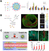
Figure 5.. Example biomaterials using proteins and enzymes present in MI for targeting.
A) Schematic of a nanoparticle containing two targeting moieties: cardiac homing peptide (yellow) and SS-31 (light blue), a mitochondrial localizing peptide. The nanoparticle was decorated with resveratrol, a therapeutic payload that acts as a reactive oxygen species scavenger. Nanoparticle localization to the infarcted LV in a rat MI model was more prevalent in co-targeting with SS-31 and the cardiac homing peptide via IVIS of whole heart images and transverse heart sections. Reproduced with permission[74]. Copyright 2019, Elsevier. B) An infusible ECM (iECM), derived from digested, decellularized myocardium, was retained in the infarcted heart in a rat MI model up to 3 days post-infusion. When infused into the heart, the iECM localized to the gaps in the leaky acute MI vasculature. Fluorescent imaging and confocal imaging both detailed endothelial cell localization (green) with iECM (red). Reproduced with permission[14]. Copyright 2022, Springer Nature Limited. C) Polynorbornene nanoparticles (NPs, black) containing a MMP responsive block (blue) formed micellular structures when in PBS. When administered in a rat MI model, MMP responsive NPs utilized the EPR effect to enter the infarct and then target via MMP upregulation, which caused a morphological switch to micron scale aggregates, in the infarcted heart. These NPs were shown to be retained in the heart up to 28 days post-injection, and required the MMP responsive block for retention. Reproduced with permission[15]. Copyright 2015, Wiley.

References

    1. Tsao CW, Aday AW, Almarzooq ZI, Alonso A, Beaton AZ, Bittencourt MS, Boehme AK, Buxton AE, Carson AP, Commodore-Mensah Y, Elkind MSV, Evenson KR, Eze-Nliam C, Ferguson JF, Generoso G, Ho JE, Kalani R, Khan SS, Kissela BM, Knutson KL, Levine DA, Lewis TT, Liu J, Loop MS, Ma J, Mussolino ME, Navaneethan SD, Perak AM, Poudel R, Rezk-Hanna M, Roth GA, Schroeder EB, Shah SH, Thacker EL, VanWagner LB, Virani SS, Voecks JH, Wang N-Y, Yaffe K, Martin SS, Circulation 2022, 145, e153. - PubMed
    1. Lu L, Liu M, Sun R, Zheng Y, Zhang P, Cell Biochem Biophys 2015, 72, 865. - PubMed
    1. Frangogiannis NG, Smith CW, Entman ML, Cardiovascular Research 2002, 53, 31. - PubMed
    1. Tous E, Purcell B, Ifkovits JL, Burdick JA, J Cardiovasc Transl Res 2011, 4, 528. - PubMed
    1. Johnson TD, Christman KL, Expert opinion on drug delivery 2013, 10, 59. - PubMed

Substances

LinkOut - more resources